


To understand how the human body moves, one must look beyond the visible surface of the skin and into the complex, hierarchical architecture of skeletal muscle. Skeletal muscle is the only tissue in the body capable of actively developing tension. This ability is not the result of a single uniform mass, but rather a highly organized system of layers and specialized fibers that work in concert to produce force, stability, and movement.
The Hierarchical Organization of Muscle Layers
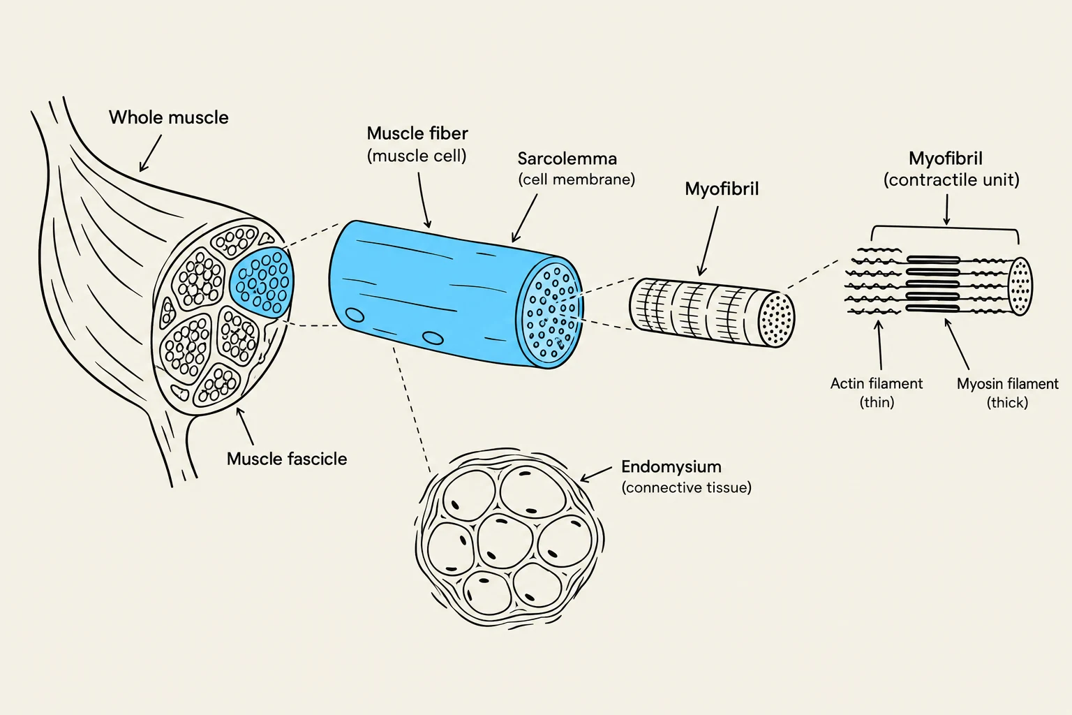
Skeletal muscle is organized into a series of nested layers, ranging from the microscopic contractile units to the macroscopic organ itself. This structural packaging ensures that force generated at the cellular level is efficiently transmitted to the bones.
At the most fundamental level, the muscle cell (also known as a muscle fiber) contains rod-like contractile units called myofibrils. These myofibrils are composed of parallel arrangements of contractile proteins—specifically actin (thin filaments) and myosin (thick filaments)—which perform the actual work of contraction. The interior of the muscle cell, which contains these components, is known as the sarcoplasm. Surrounding the individual muscle cell is a specialized plasma membrane called the sarcolemma.
Moving outward, the organization becomes more complex:
- Endomysium: The connective tissue layer that wraps around each individual muscle fiber.
- Fasciculus: A bundle of muscle fibers wrapped together.
- Perimysium: The connective tissue layer that surrounds each fasciculus.
- Epimysium: The outermost layer of connective tissue that encapsulates the entire muscle.
Keep reading for free.
Create an account to finish this reading and make it part of your study routine.
Continue reading freeAlready have an account? Log in
Updated