Understanding Epilepsy and fMRI Applications in Brain Research
This content explains epilepsy, its management with Topiramate, and the role of functional MRI (fMRI) in studying brain activity, connectivity, and clinical applications like presurgical localization. It covers task-based and resting-state fMRI, BOLD signals, and network integrity in conditions like Rolandic Epilepsy.
Epilepsy is a complex neurological disorder defined by the occurrence of recurrent seizures. These seizures are the clinical manifestation of abnormal, excessive, or synchronous electrical activity within the brain's neuronal networks. While the underlying causes can vary widely—ranging from genetic predispositions to brain injuries or infections—the primary goal of clinical management is to control these electrical disturbances to improve the patient's quality of life.
Clinical Management and the Role of Topiramate
One of the primary methods for managing epilepsy is through the use of antiepileptic drugs (AEDs). A common medication used in this context is Topiramate (TPM).
While Topiramate is highly effective at providing seizure control, it is not without complications. Clinical research often aims to examine the side effects of such treatments, specifically because TPM can lead to cognitive problems in patients. This creates a clinical challenge: balancing the necessity of seizure suppression with the preservation of cognitive function.
Functional MRI (fMRI) in Epilepsy Research
To understand how epilepsy affects the brain and how treatments like Topiramate impact cognition, researchers turn to neuroimaging. Functional Magnetic Resonance Imaging (fMRI) is a critical tool that provides a "window" into the brain's function. Unlike structural MRI, which shows anatomy, fMRI tracks brain activity by measuring the BOLD (Blood-Oxygen-Level-Dependent) signal.
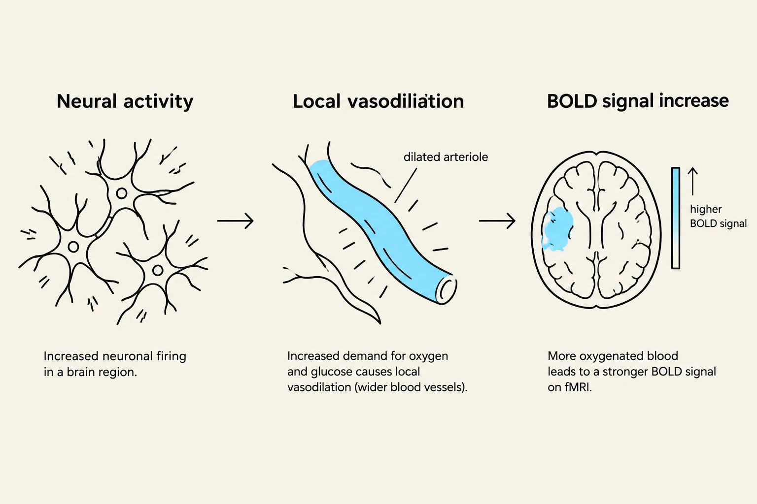
The BOLD signal relies on the relationship between oxygenated hemoglobin (O2-Hb) and deoxygenated hemoglobin (CO2-Hb). When neurons become active, there is a local increase in blood flow that "overshoots" the actual metabolic demand. This results in a higher ratio of O2-Hb to CO2-Hb, which changes the local magnetic properties of the tissue and is detected by the MRI scanner.
Task-based fMRI
In task-based fMRI, a subject performs a specific cognitive activity during the scan. If the observed neuronal signals follow the timing of the task design, researchers can identify which cortical regions are involved in that specific function.
A classic example used in epilepsy research is the word generation task. In this task, a patient is asked to generate words beginning with a specific letter (e.g., "U-N-K-A-E-P"). This task typically activates two key areas:
Broca’s Area: Responsible for expressive language.
Anterior Cingulate Cortex (ACC): Involved in executive functions.
Studies have shown that while the control group shows robust activation in these areas, the Topiramate (TPM) group may show significantly reduced activation, potentially explaining the cognitive side effects of the drug.
This diagram illustrates the process by which neural activity leads to a BOLD signal increase on fMRI. Increased neuronal firing in a brain region increases the demand for oxygen and glucose, causing local vasodilation (wider blood vessels). This results in more oxygenated blood flow, which leads to a stronger BOLD signal detected by fMRI.
Resting-state fMRI and Connectivity
Not all brain activity is tied to a specific task. Resting-state fMRI measures spontaneous BOLD signal fluctuations that occur while the patient is at rest. Interestingly, these spontaneous fluctuations—often referred to as "BOLD noise"—can account for approximately 60% of the signal.
Rather than being mere noise, these fluctuations reflect the intrinsic connectivity between different brain regions. To analyze this data without needing to pre-select specific brain regions (a priori), researchers use Independent Component Analysis (ICA). ICA is a data-driven statistical approach that decomposes the complex resting-state signal into distinct spatiotemporal patterns, allowing scientists to identify functional networks like the visual network or the default mode network.
Exam Tip: If a question asks about the difference between task-based and resting-state fMRI, remember that task-based fMRI relies on external stimuli to trigger activity, whereas resting-state fMRI relies on the spontaneous synchrony of time-series signals to map intrinsic brain networks.
Rolandic Epilepsy (RE) and Network Integrity
Rolandic Epilepsy is a specific clinical phenotype characterized by sensorimotor spikes on an EEG. Patients often present with symptoms such as hemifacial spasms, speech arrest, and language impairment.
Recent research has shifted the focus from looking at single brain regions to looking at network integrity. While the "Rolandic network" was traditionally thought to cover only the pre- and postcentral gyrus, it actually extends to include the left inferior frontal gyrus (Broca’s area).
A critical finding in RE is the breakdown of communication between two major systems:
The Motor Network
The Language Network
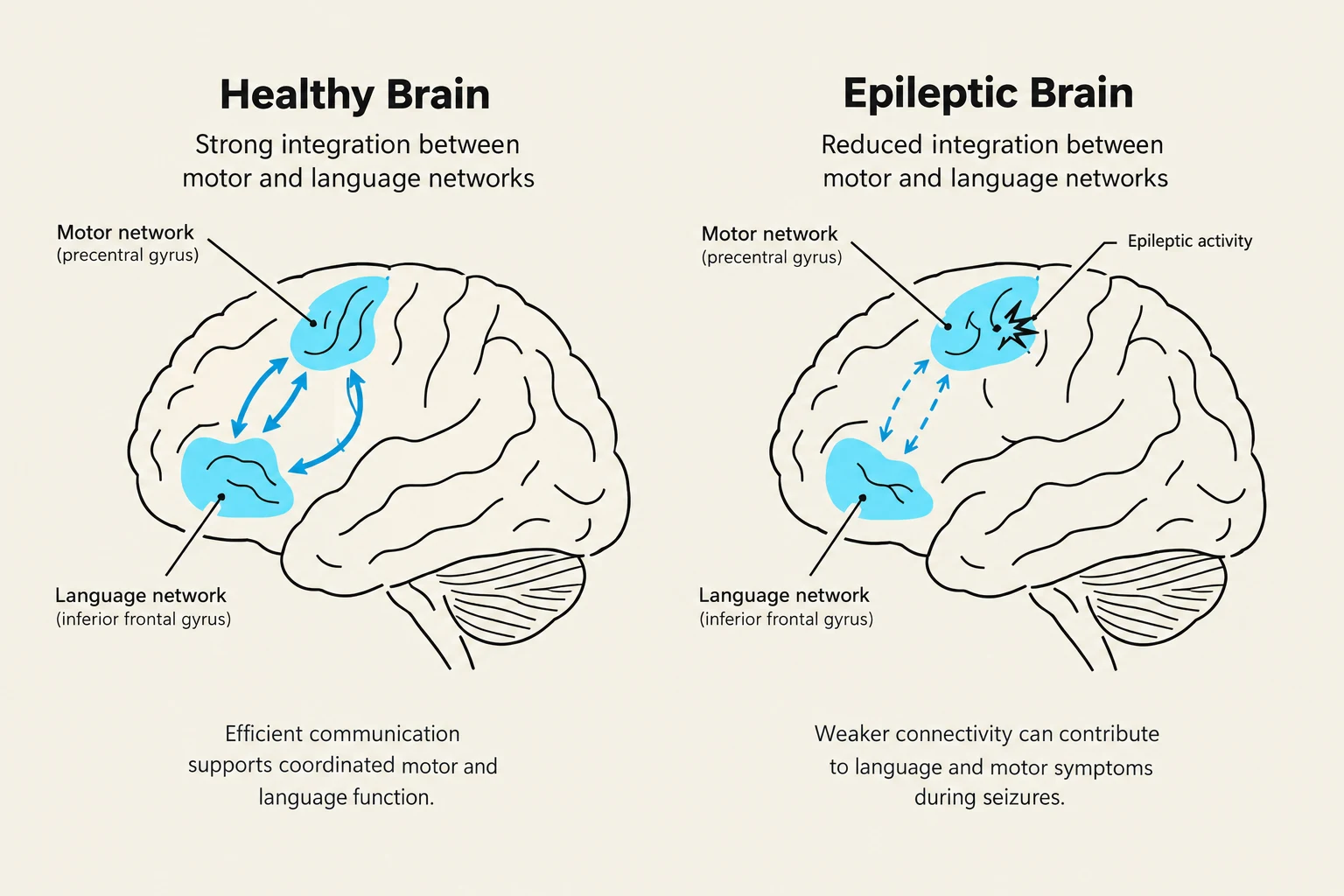
In healthy individuals, these two networks integrate at Broca's area. However, in patients with Rolandic Epilepsy, this integration is impaired, leading to reduced network integrity.
Comparison of brain networks in a healthy brain versus an epileptic brain, illustrating differences in integration and connectivity.
The Importance of Connectivity Analysis
One of the most important takeaways for students of neuroimaging is that conventional activation maps may fail to show differences in chronic epilepsy patients compared to healthy controls. If you only look at where the brain activates during a task, the epilepsy patient might look "normal."
However, when researchers apply connectivity analysis—looking at how different regions communicate over time—significant differences emerge. In chronic epilepsy, while the activation maps might appear similar, the underlying functional connectivity and the ability of the brain to integrate information across networks are significantly compromised.
Clinical Application: Presurgical Localization
Beyond research, these fMRI techniques have vital clinical applications, particularly in presurgical localization. For patients with epilepsy or tumors located near critical functional areas, surgeons must know exactly where the motor cortex (M1) is located to avoid causing permanent paralysis during surgery. By using fMRI to map the motor cortex, surgeons can use the data for precise surgical guidance, ensuring that the resection of diseased tissue does not damage essential motor or language functions.
Keep reading for free.
Create an account to finish this reading and make it part of your study routine.