Understanding Ear and Eye Disorders: Otology and Ophthalmology
An overview of common disorders affecting the ear and eye, including hearing loss, Meniere disease, conjunctivitis, cataracts, glaucoma, diabetic retinopathy, and macular degeneration. It also covers diagnostic techniques like the Weber and Rinne tests.
The sensory organs of the ear and the eyes are complex systems that allow us to navigate and interpret the world around us. When these systems are compromised by inflammation, structural changes, or degenerative processes, the impact on a patient's quality of life can be profound. This article explores the clinical landscape of otology (the study of the ear) and ophthalmology (the study of the eye), covering common disorders, diagnostic techniques, and physiological mechanisms.
Otology: Understanding Hearing and Balance
Comparison of conductive and sensorineural hearing loss mechanisms.
Hearing involves the successful transmission of sound waves from the external environment through the middle ear to the inner ear, where they are converted into electrical signals for the brain. Disorders in this pathway are generally categorized into two types: conductive and sensorineural hearing loss.
Conductive vs. Sensorineural Hearing Loss
Conductive Hearing Loss (approximately 10% of cases) occurs when there is a physical problem preventing sound waves from transferring from the outer ear to the inner ear. Common causes include:
Otitis Media: Inflammation of the middle ear, most common in children. It can cause ear pain, fever, and middle ear effusion, which may result in a bulging tympanic membrane (TM) visible during an otoscope exam.
Otosclerosis: Abnormal bone growth in the middle ear, most frequently affecting the stapes. This condition is often genetic (autosomal dominant) and most commonly affects Caucasian females in their 20s or 30s.
Other causes: Cerumen (ear wax) impaction, fluid from allergies/colds, or a perforated eardrum.
Sensorineural Hearing Loss (approximately 90% of cases) involves issues with the inner ear (cochlea) or the vestibulocochlear nerve (CN VIII). This type of loss often develops slowly and makes it difficult to hear voices against background noise. Causes include:
Presbycusis: Age-related hearing loss and the most common form of hearing loss overall. It is characterized by a progressive, bilateral decrease in hearing thresholds.
Meniere Disease: A disorder caused by fluid accumulation in the inner ear (cochlea and vestibule). It presents with a classic triad: tinnitus, vertigo (which can last 20 minutes to 24 hours), and hearing loss.
Other causes: Loud noise exposure, genetics, certain medications (e.g., aspirin, cisplatin, gentamicin), or head injuries.
Clinical Diagnosis: The Weber and Rinne Tests
The Rinne test procedure for bone and air conduction.
To differentiate between conductive and sensorineural loss, clinicians use tuning fork tests:
Weber Test (Localization): A tuning fork is placed on the center of the patient's head.
Normal: Sound is heard equally in both ears.
Abnormal: Sound is louder on one side.
Rinne Test (Conduction): A tuning fork is placed on the mastoid process and then moved near the ear canal (air conduction).
Normal: Air conduction is better than bone conduction (Air > Bone).
Sensorineural Loss: Air conduction is still better than bone conduction, but the Weber test was abnormal.
Conductive Loss: Bone conduction is better than air conduction (Bone > Air).
Exam Tip: If the Weber test shows sound is louder in one ear, immediately perform the Rinne test on that affected ear to determine if the issue is conductive or sensorineural.
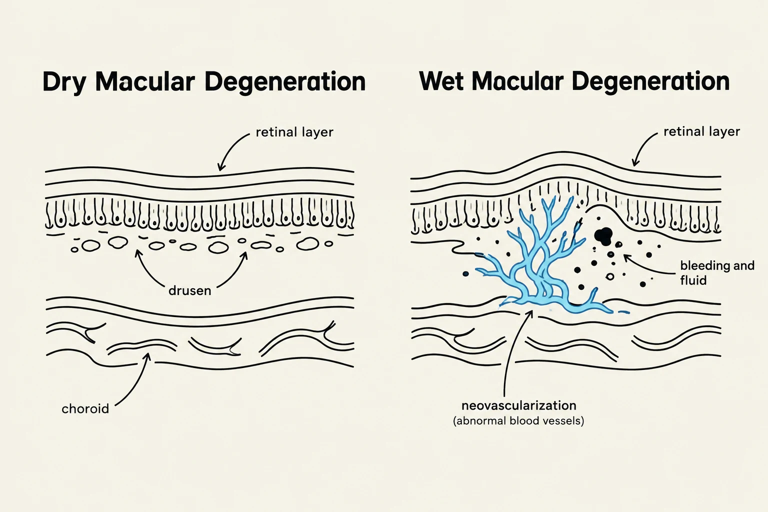
Diagrams illustrating the differences between dry and wet macular degeneration, showing the retinal layer, choroid, drusen, and neovascularization with bleeding and fluid.
Ophthalmology: External Eye and Vision Disorders
Eye health is divided into disorders affecting the external structures and those affecting the internal vision-processing mechanisms.
External Eye Disorders
The accessory structures of the eye, such as the Meibomian (tarsal) glands, provide essential lubrication.
Hordeolum (Sty): A bacterial infection of the Meibomian gland resulting in a painful, swollen eyelid. Treatment typically involves warm compresses.
Conjunctivitis (Pinkeye): Inflammation of the conjunctiva.
Bacterial: Characterized by green/yellow discharge and eyelid crusting; usually unilateral and contagious.
Viral: Characterized by watery discharge and itching; usually unilateral and contagious.
Allergic: Characterized by bilateral symptoms and is not contagious.
Vision and Internal Eye Disorders
Internal disorders can range from alignment issues in children to degenerative diseases in older adults.
Developmental and Alignment Issues:
Strabismus: Misalignment of one or both eyes (e.g., "crossed-eyes"). This can lead to headaches, double vision, or eye strain.
Amblyopia ("Lazy Eye"): Occurs when the brain and eye do not work together properly. Strabismus is the most common cause of amblyopia. Treatment, such as eye patching, must be done early in childhood to be effective.
Degenerative and Structural Disorders:
Cataracts: A cloudy or opaque discoloration of the lens, often caused by aging or UV light exposure. This leads to blurry vision and decreased color/night perception.
Glaucoma: Increased eye pressure due to fluid buildup.
Open-Angle Glaucoma: The most common type (90%); it is painless and results in a gradual loss of vision.
Closed-Angle Glaucoma: A medical emergency; it is painful, sudden, and may cause the patient to see halos around lights.
Diabetic Retinopathy: Retinal damage caused by diabetes, which is a leading cause of blindness. It can progress from Non-Proliferative (NPDR), involving microaneurysms and leaks, to Proliferative (PDR), where abnormal new blood vessels grow into the retina.
Macular Degeneration: Affects central vision in adults over 55.
Dry Macular Degeneration (90%): Slow onset; characterized by "drusen" (yellow metabolic waste deposits) under the retina.
Wet Macular Degeneration (10%): Fast onset; involves neovascularization (new blood vessel growth) that causes bleeding and irreversible damage.
Clinical Note: For many retinal conditions, including diabetic retinopathy and macular degeneration, clinicians use specialized tools like Fundoscopic Exams, Fluorescein Angiography, and Optical Coherence Tomography (OCT) for accurate diagnosis.
Keep reading for free.
Create an account to finish this reading and make it part of your study routine.