Understanding Altered Immune Responses and Hypersensitivity
This content explains altered immune responses including allergy, autoimmunity, and alloimmunity. It details the four types of hypersensitivity reactions (Type I, II, III, and IV) and their underlying mechanisms, such as IgE-mediated responses, tissue-specific reactions, immune complex formation, and cell-mediated immu
In a healthy state, the immune system maintains immunologic homeostasis, a steady state of tolerance toward the body's own antigens and a controlled lack of reaction against harmless environmental antigens. However, when this balance is disrupted, the immune system can produce responses that result in disease or damage to the host. These altered responses are broadly categorized into three types: allergy, autoimmunity, and alloimmunity.
Categories of Altered Immune Responses
Allergy
An allergy is an exaggerated immune response directed against environmental antigens (allergens). While these antigens are typically harmless, the immune system treats them as threats, leading to inflammatory symptoms.
Autoimmunity
Autoimmunity occurs when the immune response is misdirected against the host's own cells. This breakdown in self-tolerance can be driven by several mechanisms:
Exposure to sequestered antigens: Antigens that were previously hidden from the immune system are suddenly revealed.
Infectious disease complications: This often involves molecular mimicry, where a pathogen resembles host tissue.
Development of a neoantigen: Haptens may bind to self-proteins, creating a new, immunogenic target.
Ineffective peripheral tolerance: The body fails to suppress self-reactive lymphocytes.
A classic example of autoimmunity is Systemic Lupus Erythematosus (SLE), where autoantibodies attack various host components, including nucleic acids and erythrocytes, often leading to renal disease, arthritis, and malar (facial) rashes.
Alloimmunity
Alloimmunity is an immune response directed against beneficial foreign tissues, such as those encountered during blood transfusions or organ transplants.
One specific form is transient neonatal alloimmunity, such as Fetal/Neonatal Alloimmune Thrombocytopenia (FNAIT), where maternal antibodies attack fetal platelets. In the context of organ transplantation, this can lead to alloimmune graft rejection, which is categorized by its timing and mechanism into hyperacute, acute, or chronic rejection. These responses are heavily influenced by blood group antigens, specifically the ABO and Rh systems.
Understanding Hypersensitivity
Hypersensitivity refers to an altered immunologic response to an antigen that results in host damage. Two critical concepts in hypersensitivity are sensitization and desensitization. Sensitization occurs when an individual develops an adequate amount of antibodies or T cells so that a noticeable reaction occurs upon reexposure to the antigen. Desensitization is the clinical process of reducing this sensitivity.
Hypersensitivity reactions are also classified by their timing:
Immediate hypersensitivity: Occurs within minutes to a few hours after exposure.
Delayed hypersensitivity: Takes several hours to appear, reaching maximum severity days after reexposure.
Exam Tip: When distinguishing between hypersensitivity types, always look at whether the mediator is an antibody (Types I, II, III) or a cell (Type IV).
The Four Types of Hypersensitivity
Type I: Immediate (IgE-Mediated)
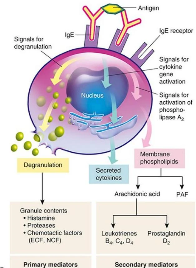
Type I hypersensitivity is mediated by Immunoglobulin E (IgE) against environmental allergens. When an allergen enters the body, IgE binds to receptors on the surface of mast cells. Upon reexposure, the antigen cross-links these IgE molecules, triggering mast cell degranulation and the release of mediators like histamine.
Diagram illustrating the process of mast cell degranulation and the release of mediators in response to antigen binding to IgE receptors.
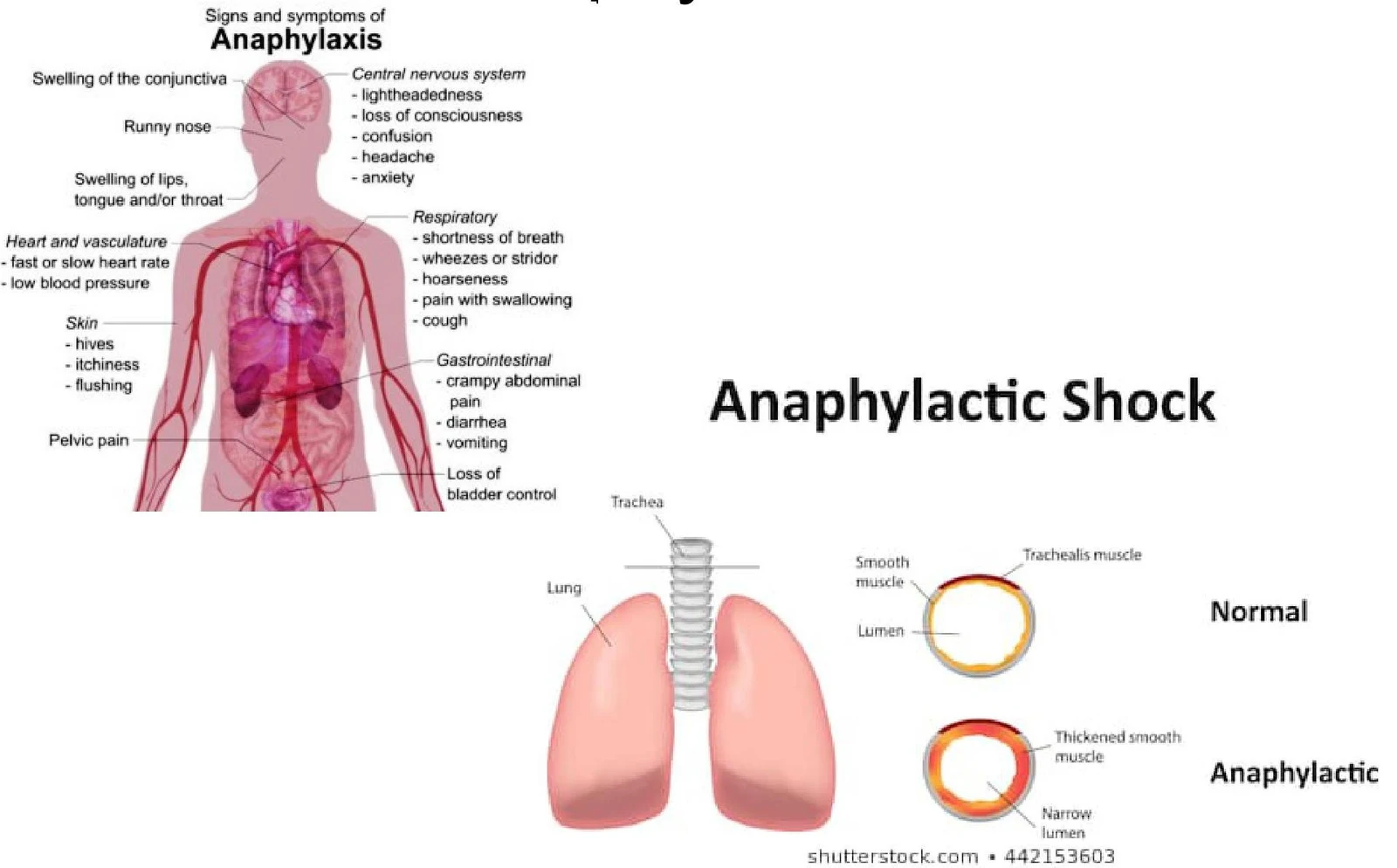
This release increases chemotactic activity and can lead to various clinical symptoms, including hives, swelling, and respiratory distress. In severe cases, this results in anaphylaxis, a systemic reaction characterized by hypotension (low blood pressure), tachycardia (fast heart rate), swelling of the throat, and potential loss of consciousness.
This illustration depicts various symptoms and internal effects of anaphylaxis, including skin reactions like urticaria and angioedema, respiratory issues such as bronchospasm and laryngeal edema, gastrointestinal distress, and cardiovascular changes like hypotension.
This illustration details the signs and symptoms of anaphylaxis, including effects on the central nervous system, respiratory system, gastrointestinal system, skin, heart, vasculature, and pelvic region. It also shows a comparison of a normal trachea cross-section with one affected by anaphylaxis, illustrating the narrowing of the airway due to smooth muscle thickening.
Type II: Tissue-Specific
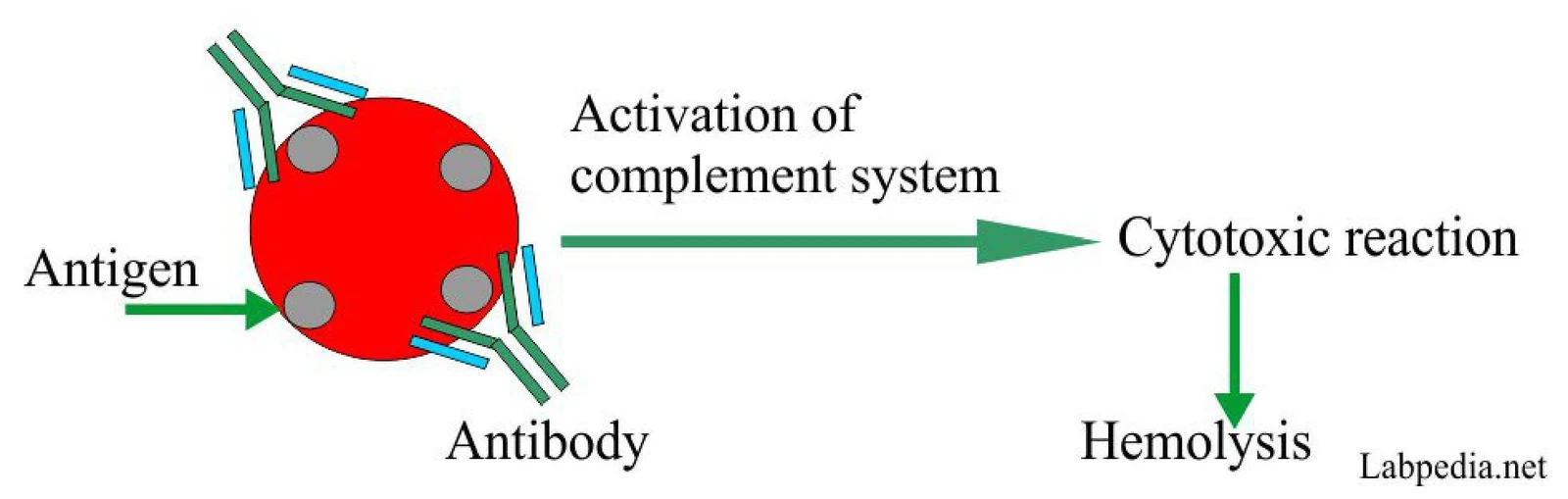
Type II hypersensitivity targets specific cells or tissues via tissue-specific antigens. Damage occurs through several mechanisms, including complement activation, phagocytosis, neutrophil granule release, or antibody-dependent cell-mediated cytotoxicity. A common clinical example is a blood transfusion reaction, where antibodies trigger the complement system, leading to the hemolysis (destruction) of red blood cells.
Diagram illustrating the complement system activation leading to a cytotoxic reaction and hemolysis.
Type III: Immune Complex-Mediated
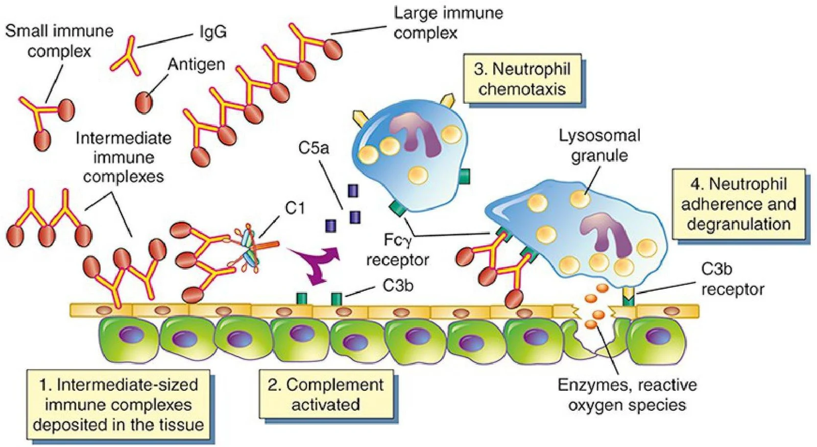
Type III hypersensitivity involves the formation of antigen-antibody complexes in the circulation. These complexes are not organ-specific but deposit in vessel walls or extravascular tissues (such as the glomerulus in the kidneys). Damage is caused by complement activation and the release of neutrophil lysosomal enzymes.
The severity of the reaction depends on the size of the complexes:
Large complexes: Cleared by macrophages.
Small complexes: Cleared via renal clearance.
Intermediate complexes: These are the most problematic because they deposit in tissues, causing damage.
This diagram illustrates the process of immune complex formation and the subsequent activation of neutrophils. It shows how intermediate-sized immune complexes deposited in tissue can activate the complement system, leading to neutrophil chemotaxis, adherence, and degranulation. The diagram labels various components involved, including antigens, IgG antibodies, complement factors (C1, C3b, C5a), Fcγ receptors, and lysosomes within neutrophils.
Type IV: Cell-Mediated (Delayed)
Unlike the first three types, Type IV hypersensitivity is cell-mediated and does not involve antibodies. Instead, it is driven by T lymphocytes. Tissue destruction occurs through two primary pathways:
Cytotoxic T (Tc) cells: These cells cause direct killing of target cells via the release of toxins.
Helper T (Th) cells: Specifically, Th1 and Th17 cells produce cytokines that recruit phagocytes, such as macrophages, to the site of the antigen.
This diagram illustrates the mechanisms of cell-mediated immunity, showing how cytotoxic T cells (Tc cells) induce apoptosis in target cells via Fas ligand interaction and the release of granzymes and perforin. It also depicts the role of Th1 cells in activating macrophages through IFN-gamma signaling, leading to the release of lysosomal enzymes and toxic oxygen species.
Keep reading for free.
Create an account to finish this reading and make it part of your study routine.