Skeletal and Passive Tissues: Stability, Lubrication, and Load Management
An overview of the musculoskeletal system, focusing on passive tissues like ligaments, tendons, and cartilage. It explains mechanical loads, stress, strain, and the roles these tissues play in joint stability, lubrication, and force distribution.
The human musculoskeletal system is a marvel of engineering, designed to facilitate movement while simultaneously protecting the body from the destructive potential of mechanical loads. While bones provide the structural framework, they cannot function in isolation. To prevent bones from breaking one another or dislocating during movement, the body relies on a specialized group of "passive tissues". These include ligaments, tendons, and various forms of cartilage, which work together to manage friction, absorb shock, and provide the necessary structural stability to withstand complex physical stresses.
Understanding Mechanical Loads and Stress
Before examining specific tissues, it is essential to understand the forces they must resist. Mechanical loads can be categorized as "pushes" or "pulls" and can be either contact or non-contact forces. When these forces act upon a tissue, they can set it in motion, change its velocity, stop its motion, change its direction, or even change its shape.
Tissues are subjected to several distinct types of loading:
Compression: A "squishing" force acting along the longitudinal axis, such as the weight of the body pressing down on the vertebrae.
Tension: A "pulling apart" force, often produced by muscles pulling on bone attachments.
Shear: A "sliding" force where the load acts parallel to the tissue surface, often a combination of compression and tension.
Torsion: A twisting force acting along the longitudinal axis, which can lead to spiral fractures.
The impact of these loads is defined by stress and strain. Stress is the manner in which a force is distributed over a specific area, calculated as:
Stress=AreaForce
Strain refers to the resulting deformation or change in shape of the tissue. The relationship between these two determines a tissue's stiffness, which is the ratio of stress to strain:
Stiffness=StrainStress
Exam Tip: Remember that bone is the stiffest tissue in the human body, while skin has the least stiffness. Tendons, ligaments, and cartilage fall somewhere in the middle.
Ligaments and Tendons: The Connectors
Passive tissues are primarily categorized by what they connect.
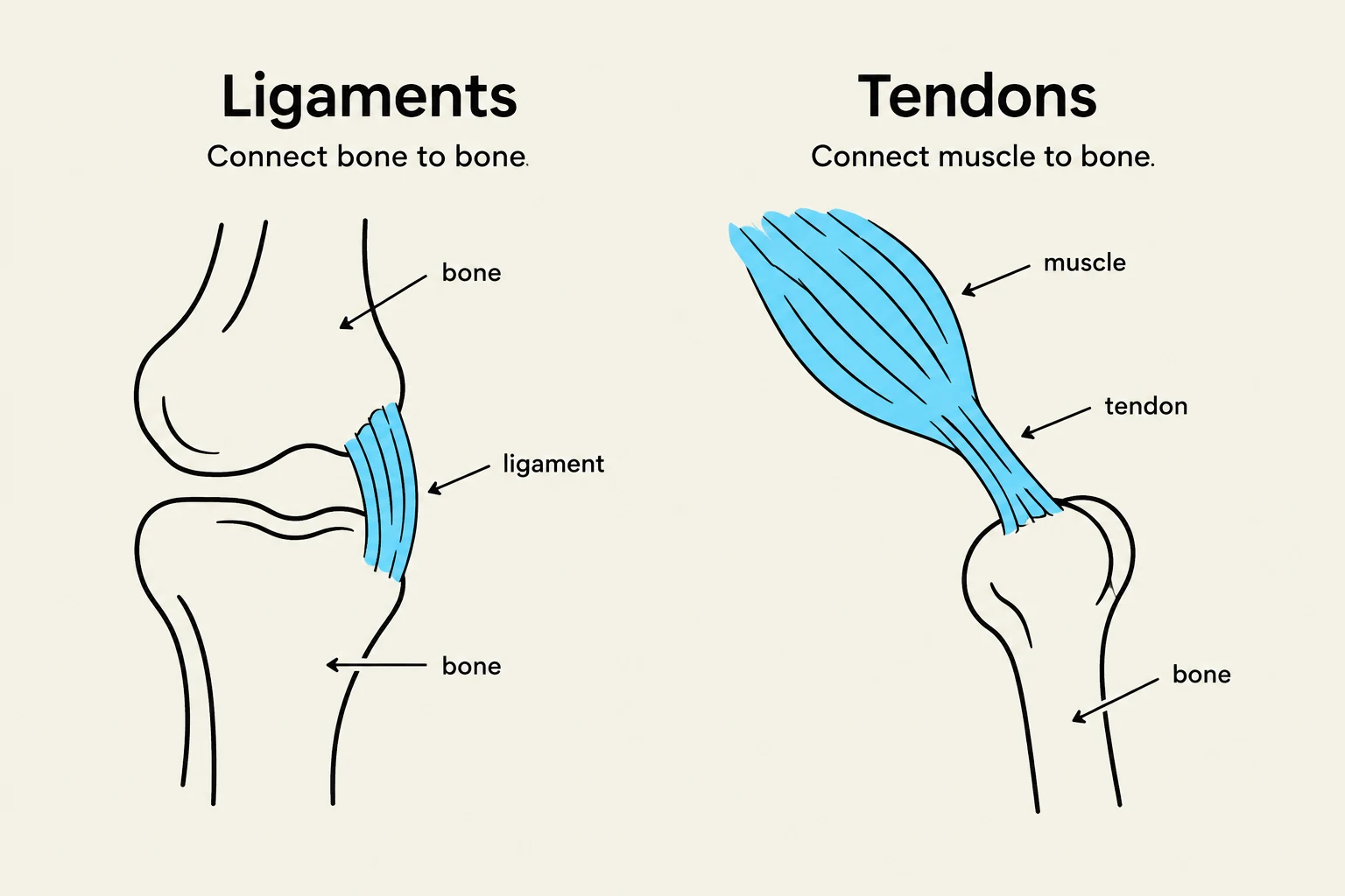
Ligaments
Ligaments are dense connective tissues that join bone to bone. They function as passive joint stabilizers, preventing excessive movement or dislocations. A ligament is composed of a complex matrix of ground substance and is approximately 70% collagen. These collagen bundles are organized specifically to withstand the biomechanical stresses typical of that joint.
Clinically, when a ligament is injured, the condition is classified as a sprain. Ligaments are often named based on their specific characteristics, such as their bony attachment (e.g., coracoacromial), their shape (e.g., deltoid), or their relationship to other ligaments (e.g., cruciate).
Tendons
While similar in composition to ligaments, tendons serve a different mechanical purpose: they connect muscle to bone. They are essential for transmitting the force generated by muscle contraction to the skeletal system to initiate movement.
Diagrams illustrating the difference between ligaments, which connect bone to bone, and tendons, which connect muscle to bone.
Articular Connective Tissue: Cartilage
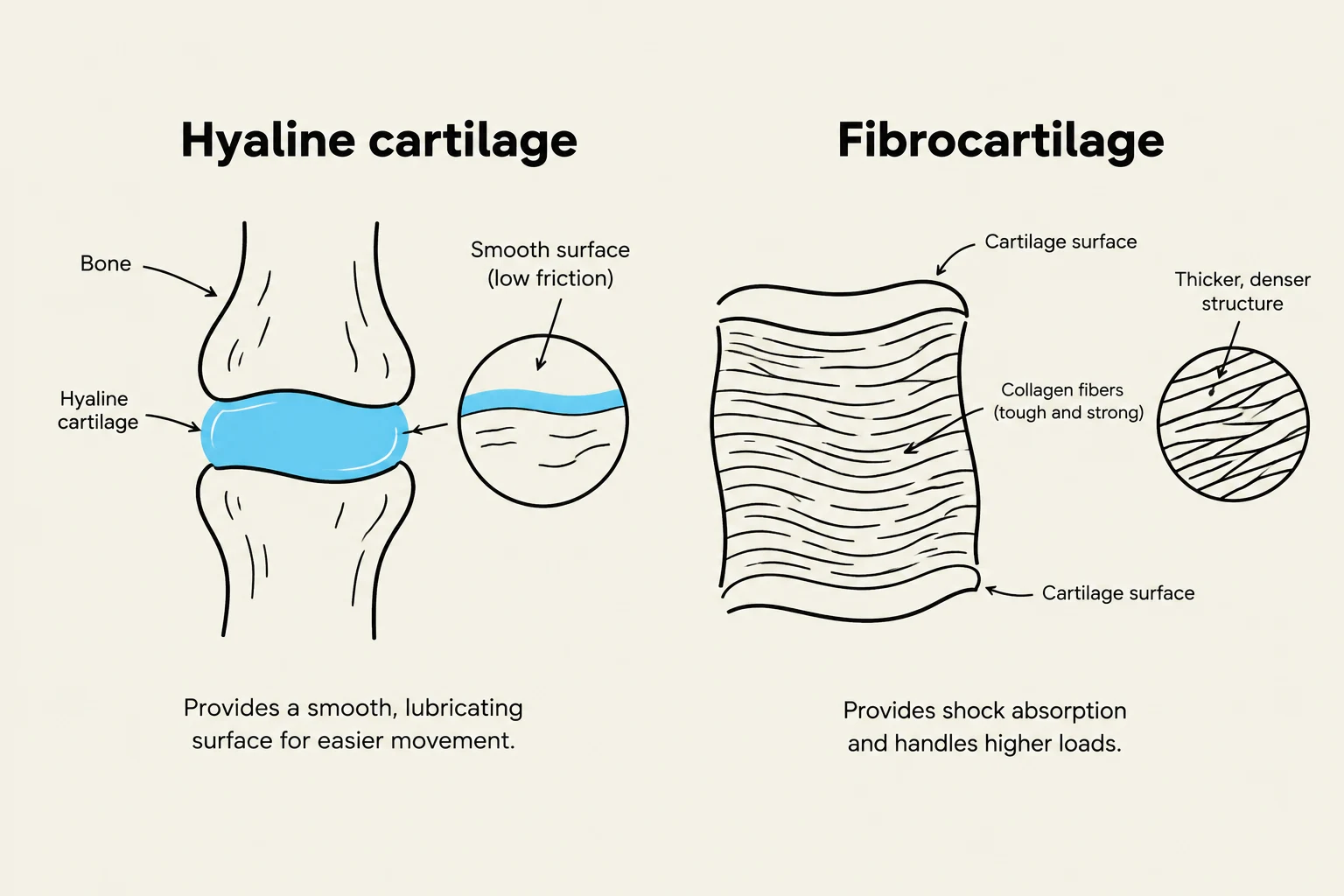
Articular tissues are specialized to protect diarthrodial (freely movable) joints by managing friction and distributing loads. There are two primary types of cartilage used in the body: hyaline and fibrocartilage.
Hyaline Cartilage
Hyaline cartilage is a soft, porous, and highly hydrated dense white tissue that covers the ends of bones in diarthrodial joints. It is maintained by specialized cells called chondrocytes, which make up less than 10% of the tissue's volume. These cells manage a matrix composed of collagen, proteins, and water.
The primary roles of hyaline cartilage are to reduce friction between bone ends and to spread force over wide areas to decrease localized stress. It achieves this through a unique mechanism known as weeping lubrication:
When the joint is loaded, the cartilage deforms.
This deformation causes synovial fluid to "flow" out of the porous matrix.
At low loading rates, the solid matrix itself resists the load.
At high loading rates, the fluid within the matrix resists the load, which can reduce contact stress by up to 50%.
Fibrocartilage
Fibrocartilage is a tougher, more resilient tissue designed for high-stress environments. It is found in specialized structures such as the intervertebral discs of the spine and the menisci of the knee.
Unlike the smooth surface of hyaline cartilage, fibrocartilage is optimized for:
Enhanced shock absorption.
Improving the "fit" between articulating bone surfaces.
Limiting excessive bone movement and further distributing force across the joint.
Diagrams illustrating the structure and function of hyaline cartilage and fibrocartilage.
Joint Stability and Architecture
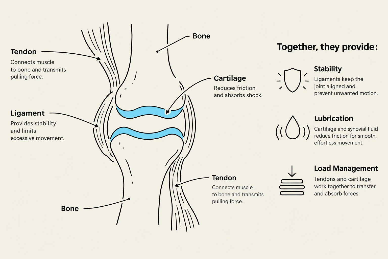
The effectiveness of these passive tissues is heavily influenced by the geometry of the bones they support. Joint stability refers to a joint's ability to resist dislocation. This stability is a product of the shape of the articulating surfaces and the supporting soft tissues (ligaments, tendons, and cartilage).
For example, the hip joint has a deep socket, which increases the contact surface area and provides high stability. In contrast, the shoulder joint has a much shallower socket, resulting in a smaller contact area and greater mobility but less inherent stability.
Key Concept: Joints can exist in a "Close-Packed Position," where the articulating surfaces have their best fit and maximum contact, providing the greatest stability. Any other position is considered "Loose-Packed" and is less stable.
Summary of Tissue Roles
To maintain a functional and pain-free musculoskeletal system, these tissues must work in harmony to manage the constant cycle of loading and unloading.
Tissue Type
Primary Connection/Location
Main Function
Ligament
Bone to Bone
Passive stability; resists dislocation
Tendon
Muscle to Bone
Transmits muscle force to bone
Hyaline Cartilage
Articular surfaces
Lubrication; force distribution
Fibrocartilage
Menisci; Intervertebral discs
Shock absorption; improves joint fit
This diagram illustrates the anatomy of a joint, detailing the roles of bones, tendons, ligaments, and cartilage in providing stability, lubrication, and load management.
Keep reading for free.
Create an account to finish this reading and make it part of your study routine.