An explanation of metabolic syndrome, its diagnostic criteria, and the pathophysiological mechanisms involving adipose tissue, insulin resistance, and inflammation. Covers clinical complications like type 2 diabetes and cardiovascular disease.
Metabolic syndrome is not a single disease entity, but rather a dangerous clustering of interconnected metabolic abnormalities. When these conditions co-occur, they significantly amplify the risk of severe systemic complications, including cardiovascular disease and diabetes. Understanding the transition from simple weight gain to complex metabolic dysfunction requires an exploration of how adipose tissue behaves as an endocrine organ and how its failure drives systemic insulin resistance.
Defining Metabolic Syndrome and Obesity
Clinically, metabolic syndrome is diagnosed when a patient presents with at least three of the following five criteria:
Abdominal obesity: Waist circumference ≥102 cm in men or ≥88 cm in women.
Low HDL cholesterol: ≤40 mg/dL in men or ≤50 mg/dL in women.
Obesity is the primary driver of this cluster and is clinically assessed using the Body Mass Index (BMI). BMI is calculated using the following formula:
BMI=height (m)2weight (kg)
According to standard classifications, a BMI ≥30.0 kg/m2 is considered obese, while a BMI >40.0 kg/m2 is classified as morbid obesity.
Exam Tip: Note the distinction between clinical obesity (based on BMI) and the conceptual definition (excess body fat). A patient can have a high BMI, but the location of the fat is often more predictive of metabolic risk than the weight itself.
The Role of Adipose Tissue: Subcutaneous vs. Visceral
Not all fat is created equal. There is a critical physiological distinction between subcutaneous fat, which is stored just beneath the skin, and visceral fat, which is found deep within the abdominal cavity surrounding vital organs like the liver and intestines.
Visceral adiposity is a primary driver of metabolic dysfunction. While subcutaneous fat acts largely as an energy reservoir, visceral fat is highly metabolically active and contributes significantly to the systemic inflammatory state and insulin resistance.
Pathophysiology: Hypertrophy, Hyperplasia, and Inflammation
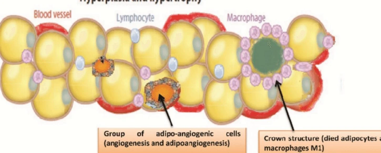
As energy intake exceeds expenditure, adipose tissue expands through two primary mechanisms: hyperplasia (an increase in the number of adipocytes) and hypertrophy (an increase in the size of existing adipocytes).
In a healthy, "lean" state, adipose tissue maintains adequate blood supply and contains anti-inflammatory immune cells. However, as obesity progresses, the tissue undergoes pathological changes:
Adipocyte Hypertrophy: As cells enlarge, they experience mechanical stress and may outgrow their blood supply, leading to hypoxia (low oxygen).
Necrosis and Inflammation: Hypoxic, stressed cells may undergo necrosis (cell death). This triggers an immune response where macrophages infiltrate the tissue.
Cytokine Release: These inflammatory cells and stressed adipocytes release pro-inflammatory cytokines (adipokines), specifically TNF-α and IL-6.
Diagram illustrating adipocytes, lymphocytes, macrophages, and blood vessels, with labels indicating a group of adipo-angiogenic cells and a crown structure formed by died adipocytes and macrophages M1.
This chronic, low-grade inflammation, combined with reduced capillary density and increased collagen deposition, transforms the adipose tissue into a dysfunctional organ that actively promotes metabolic disease.
The Central Mechanism: Insulin Resistance
The transition from obesity to metabolic syndrome is mediated largely by insulin resistance. In a healthy physiological state, insulin acts to lower blood glucose by promoting uptake in the muscles and suppressing glucose production in the liver.
In a state of insulin resistance, the signaling pathway is disrupted across multiple tissues:
Skeletal Muscle: There is a decreased ability to take up glucose from the blood.
Liver: Instead of suppressing glucose production, the liver increases it, while simultaneously increasing the synthesis of VLDL and triglycerides.
Adipose Tissue: Insulin fails to suppress lipolysis (the breakdown of fats). This causes a massive flux of free fatty acids (FFAs) into the bloodstream.
The resulting cascade leads to several hallmark pathological states:
Hyperinsulinemia: The pancreas overproduces insulin to compensate for resistance.
Hyperglycemia: Despite high insulin levels, blood glucose remains elevated because the cells cannot utilize it.
Dyslipidemia: Characterized by high triglycerides and low HDL cholesterol.
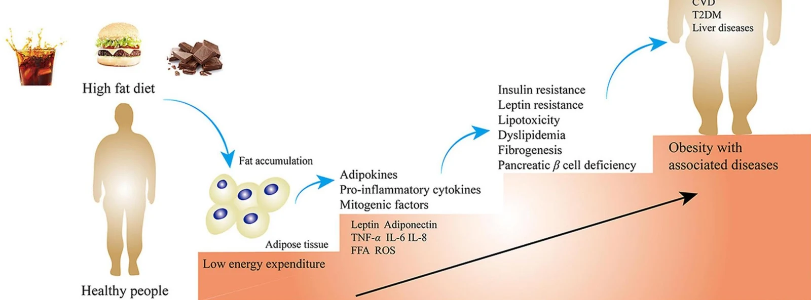
This diagram illustrates the progression from a high-fat diet and low energy expenditure to fat accumulation in adipose tissue, leading to the release of adipokines and pro-inflammatory cytokines. This cascade can result in insulin resistance, lipotoxicity, dyslipidemia, and pancreatic beta cell deficiency, ultimately contributing to obesity with associated diseases such as cardiovascular disease (CVD), type 2 diabetes mellitus (T2DM), and liver diseases.
Clinical Complications and Comorbidities
The systemic effects of insulin resistance, FFAs, and chronic inflammation extend far beyond adipose tissue. The combination of endothelial dysfunction (damage to blood vessel linings), neurohormonal activation, and oxidative stress creates a high-risk environment for multiple end-organ diseases:
Type 2 Diabetes: Driven by the failure of pancreatic beta cells to overcome insulin resistance and chronic hyperglycemia.
Atherosclerotic Cardiovascular Disease (ASCVD): Dyslipidemia and inflammation accelerate plaque formation in the arteries, increasing the risk of stroke and myocardial infarction.
Nonalcoholic Fatty Liver Disease (NAFLD): Driven by the influx of FFAs to the liver and increased hepatic lipid synthesis.
Chronic Kidney Disease (CKD): Resulting from the combined effects of hypertension and hyperglycemia on the renal microvasculature.
Management of these conditions focuses heavily on lifestyle interventions—addressing physical inactivity and excessive sugar consumption—to mitigate the primary risk factors of obesity and metabolic syndrome.
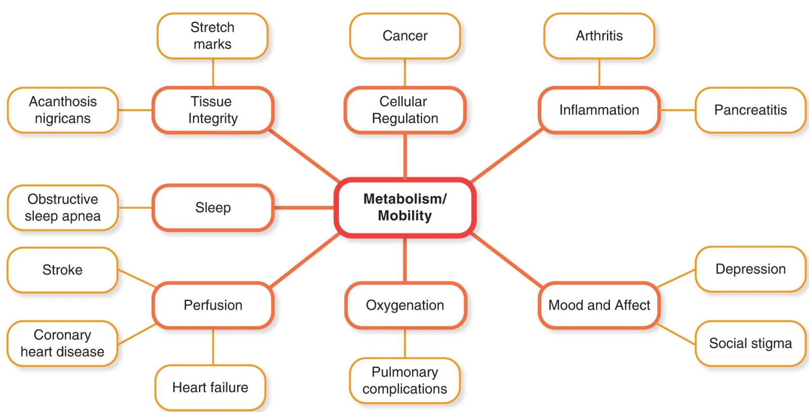
This diagram illustrates the interconnectedness of metabolism and mobility with various physiological and psychological conditions, including tissue integrity, sleep, perfusion, oxygenation, cellular regulation, inflammation, and mood and affect.
Keep reading for free.
Create an account to finish this reading and make it part of your study routine.