This content explores the components of innate resistance, the physiological pathways of inflammation, and the roles of various cytokines and phagocytes in the body's defense system.
When a pathogen enters the body or tissue is damaged, the body does not wait for a specialized response to develop. Instead, it relies on a rapid, non-specific defense system known as innate immunity. This system acts as the immediate first line of defense, utilizing physical, chemical, and cellular barriers to prevent infection and initiate the healing process.
Innate Resistance vs. Adaptive Immunity
The immune response is generally divided into two main categories: innate immunity and adaptive (acquired) immunity.
Innate resistance is characterized by its immediacy; it responds within seconds to minutes. It is non-specific, meaning it recognizes general patterns common to many pathogens rather than specific individual strains. The first line of defense includes physical barriers like the skin and mucus, as well as chemical secretions such as sweat, saliva, tears, and earwax. These secretions often contain lysozymes and antimicrobial peptides like defensins and cathelicidins, which work by disrupting bacterial cell membranes.
In contrast, adaptive immunity is a more specialized "third line of defense". While it takes longer to activate, it possesses "memory," allowing the body to recognize and respond more effectively to a pathogen it has encountered before.
This diagram illustrates the key components and processes of innate and adaptive immunity, including the roles of various cells and molecules in defending the body against pathogens.
The Inflammatory Response and its Cardinal Signs
When innate barriers are breached by injury or infection, the body initiates inflammation. Inflammation is a complex biological response intended to localize the injury, eliminate the cause, and prepare the tissue for repair.
Clinically, acute inflammation is identified by five "cardinal signs":
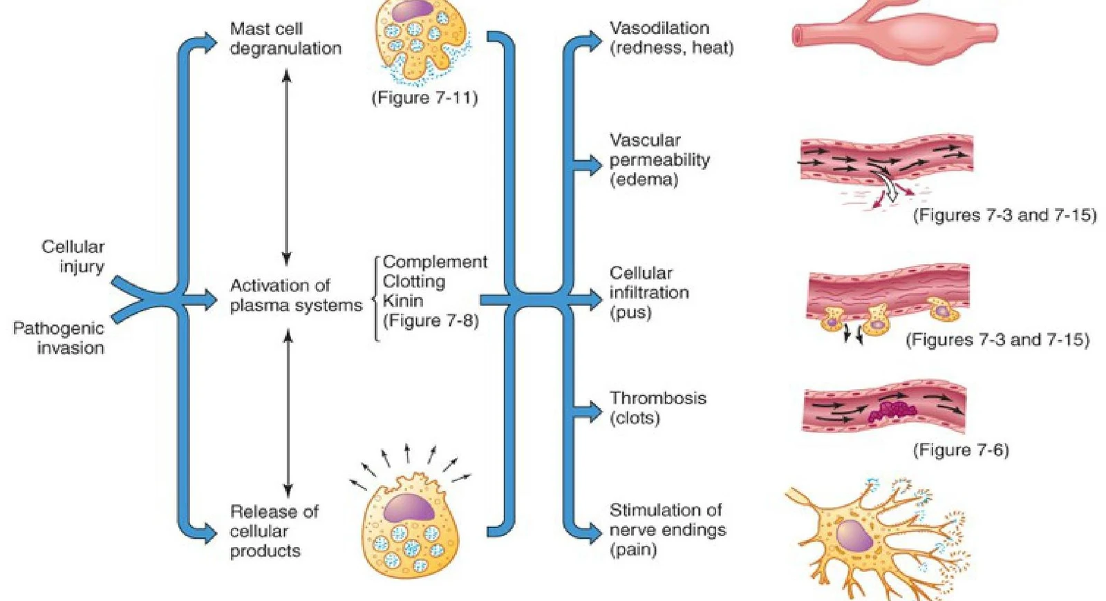
Rubor (redness): Caused by increased blood flow (vasodilation).
Tumor (swelling): Caused by increased vascular permeability and fluid accumulation.
Calor (heat): Resulting from increased blood flow to the area.
Dolor (pain): Triggered by chemical mediators and pressure on nerve endings.
Functio laesa (loss of function): A later addition to the classic signs, representing the impact of injury on the affected part.
This diagram illustrates the inflammatory response, showing how cellular injury or pathogenic invasion triggers plasma system activation, leading to mast cell degranulation and the release of cellular products. These processes result in vasodilation, increased vascular permeability, cellular infiltration, thrombosis, and stimulation of nerve endings, causing pain.
Plasma Protein Systems: Complement, Coagulation, and Kinin
The inflammatory process is driven by three interconnected plasma protein systems that work in concert to protect the body:
Complement System: This system consists of proteins that, when activated via the classical, lectin, or alternative pathways, facilitate cell lysis (destroying the pathogen) and opsonization (tagging pathogens for destruction).
Coagulation System: This system is responsible for blood clotting, which helps wall off the injured area and prevents the spread of pathogens.
Kinin System: This system produces kinins, which are potent mediators that contribute to vascular changes (vasodilation) and the sensation of pain.
Cellular Recognition: PRRs, PAMPs, and DAMPs
How do immune cells "know" there is an intruder? The bridge between innate and adaptive immunity lies in Pattern Recognition Receptors (PRRs). These receptors are expressed on the surface of immune cells to detect specific molecular signatures:
PAMPs (Pathogen-Associated Molecular Patterns): Molecules common to many microbes, such as bacterial cell wall components.
DAMPs (Damage-Associated Molecular Patterns): Molecules released by our own stressed or dying cells during injury.
Key types of PRRs include Toll-like receptors (TLRs), which recognize viral proteins and cell wall components, as well as complement receptors and scavenger receptors on macrophages that facilitate the engulfment of pathogens.
Chemical Mediators: Mast Cells and Lipid Products
Once a threat is recognized, cells like mast cells undergo degranulation, releasing preformed biochemical mediators into the surrounding tissue.
Histamine and its Receptors
One of the most important preformed mediators is histamine. Histamine binds to specific receptors to trigger different physiological effects:
H1 Receptors: Mediate smooth muscle contraction and increase vascular permeability by causing endothelial cells to contract (creating gaps in the vessel wall).
H2 Receptors: Primarily involved in stimulating gastric acid secretion in the stomach.
Lipid Mediators: Prostaglandins and Leukotrienes
In addition to releasing preformed granules, mast cells synthesize new lipid mediators from arachidonic acid:
Prostaglandins: These drive vasodilation and contribute significantly to the sensation of pain.
Leukotrienes: These are potent mediators of vascular permeability and can trigger bronchoconstriction.
Phagocytes: The Cellular Workhorses
Phagocytes are specialized cells that ingest and destroy pathogens through a process called phagocytosis.
Neutrophils
Neutrophils are typically the "first responders" to an acute inflammatory site. They migrate toward the injury via chemotaxis (movement guided by chemical signals like chemokines). To destroy engulfed bacteria, neutrophils utilize the respiratory burst, a process where the enzyme NADPH oxidase generates reactive oxygen species to kill the pathogen. Neutrophils can also employ NETosis, where they release "neutrophil extracellular traps" (NETs) to ensnare and kill microbes.
Macrophages
Macrophages arrive later and play a more versatile role. They can polarize into different phenotypes based on the environment:
M1 Phenotype: Pro-inflammatory; these cells actively fight infection and release cytokines.
M2 Phenotype: Anti-inflammatory; these cells focus on tissue repair and resolution.
Cytokines and Systemic Effects
Cells communicate through signaling proteins called cytokines.
Exam Tip: Distinguish between the roles of pro-inflammatory and anti-inflammatory cytokines. Pro-inflammatory cytokines (like TNF-α, IL-1, and IL-6) drive the response, while anti-inflammatory cytokines (like IL-10 and TGF-β) limit it.
Pro-inflammatory cytokines (TNF-α, IL-1, IL-6) can cause systemic effects, including:
Fever: Triggered by the hypothalamus in response to cytokines.
Leukocytosis: An increase in the white blood cell count in the blood.
Acute-phase protein synthesis: The liver increases production of proteins like C-reactive protein (CRP).
Anti-inflammatory cytokines (IL-10, TGF-β) are essential for limiting the inflammatory response and promoting tissue resolution or repair.
Acute vs. Chronic Inflammation and Outcomes
Inflammation is categorized by its duration and the nature of the stimulus:
Feature
Acute Inflammation
Chronic Inflammation
Onset
Immediate
Delayed
Duration
Few days
Months to years
Major Cells
Neutrophils, Basophils, Eosinophils
Monocytes, Macrophages, Lymphocytes
Outcome
Resolution, abscess, or chronic inflammation
Tissue destruction, fibrosis
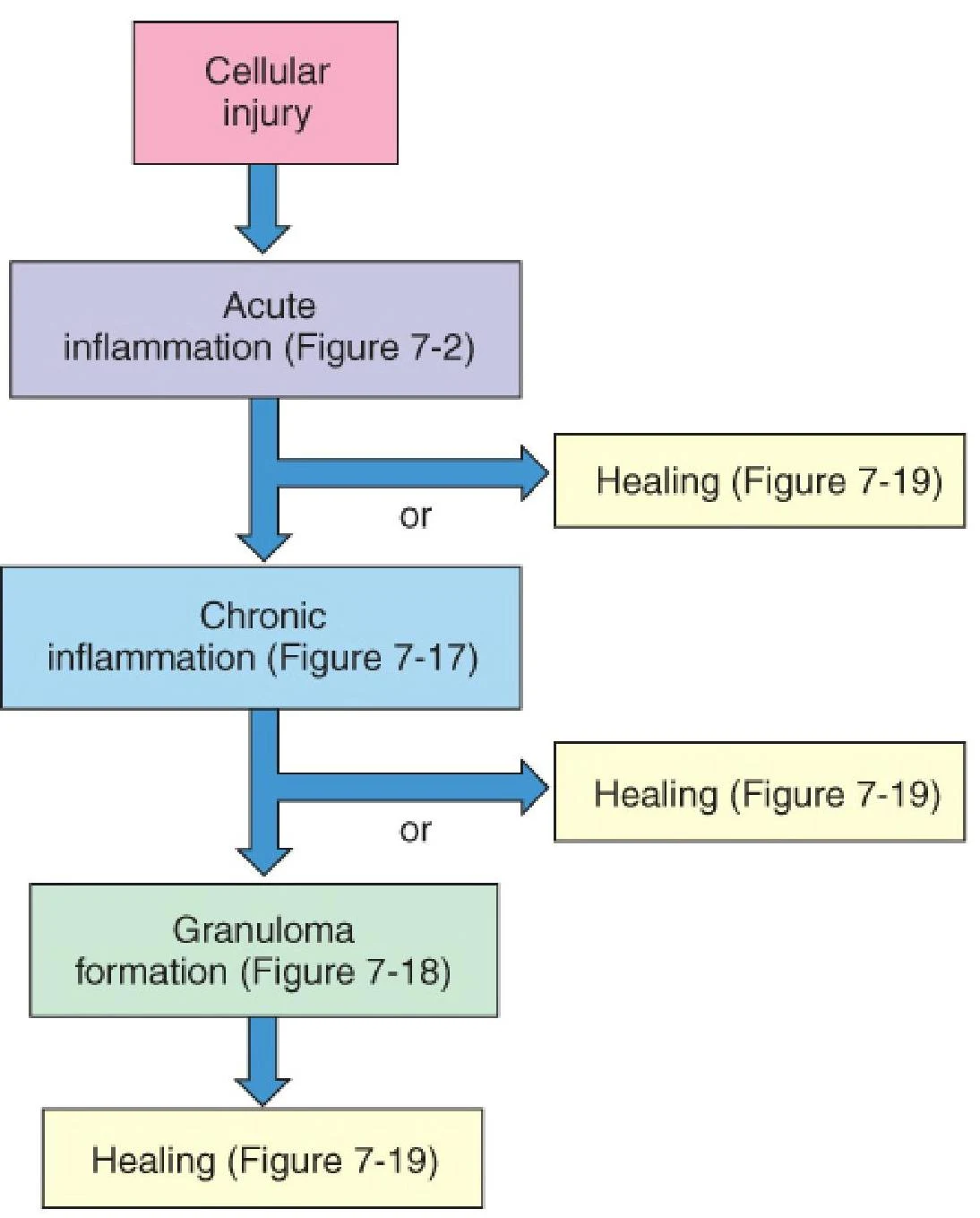
In cases of chronic inflammation, where a stimulus (like a non-degradable foreign body or an autoimmune reaction) persists, the body may form a granuloma. A granuloma is a specialized cluster of immune cells, including epithelioid macrophages and lymphocytes, that attempts to wall off the persistent irritant.
The Final Result: Resolution or Repair
The ultimate goal of the inflammatory process is to return the tissue to homeostasis:
Tissue Resolution: The complete restoration of normal tissue structure.
Repair: When damage is too extensive for resolution, the body replaces damaged tissue with connective tissue, resulting in scarring.
This diagram illustrates the potential outcomes of cellular injury, including acute inflammation, chronic inflammation, and granuloma formation, all of which can lead to healing.
Keep reading for free.
Create an account to finish this reading and make it part of your study routine.