Joint Architecture and Stability in Human Movement
Explores the fundamental principles of joint stability, including the roles of articulating surface geometry and supporting soft tissues. Discusses mechanical positions (close-packed vs. loose-packed) and classification systems for joints based on movement and architecture.
In the study of human movement, the joint is the fundamental unit where structure meets function. A joint is not merely a gap between two bones; it is a complex integration of bone, ligaments, capsules, cartilage, and muscles. This biological assembly governs our ability to move, providing the necessary mechanics for everything from the delicate rotation of a finger to the powerful weight-bearing capacity of the hip. Understanding how these components work together is essential to understanding how the body maintains its integrity under load and how it resists injury.
Understanding Joint Stability and Dislocation
The primary mechanical goal of a joint is to maintain joint stability. Stability is defined as the capacity of a joint to resist dislocation. A dislocation occurs when the articulating surfaces of a joint are forced out of their normal alignment, a state that can lead to significant damage to the surrounding bones, ligaments, tendons, and muscles.
Stability is not a static property; it is a dynamic balance between the geometry of the bones and the tension provided by the surrounding soft tissues. While the bones provide the foundational structural fit, the tissues crossing the joint—such as muscles and ligaments—provide the active and passive support necessary to maintain alignment during movement.
Exam Tip: When assessing stability, remember that it is a trade-off. Generally, the more stable a joint is (like the hip), the less mobile it becomes. Conversely, the more mobile a joint is (like the shoulder), the less inherently stable it is.
The Two Pillars of Stability: Geometry and Soft Tissue
The stability of any given joint is primarily determined by two factors: the geometry of the articulating surfaces and the supporting soft tissues.
1. Articulating Surface Geometry
The shape of the bones—specifically how the "male" and "female" parts of the joint fit together—dictates the contact area. A larger contact area generally leads to higher stability because the force is distributed more effectively across the surface.
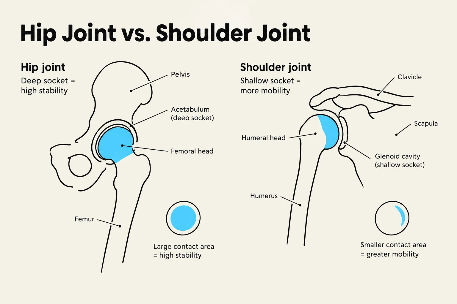
Deep Sockets: A joint with a deep socket, such as the hip, increases the contact surface area, which enhances stability.
Shallow Sockets: A joint with a shallow socket, such as the shoulder, decreases the contact area, which reduces stability but allows for a much greater range of motion.
Comparison of the hip joint and shoulder joint, illustrating differences in socket depth, contact area, and resulting stability versus mobility.
2. Supporting Soft Tissues
Even a perfectly shaped bone cannot remain stable without help. The surrounding tissues act as the "reinforcements" of the joint:
Ligaments: These are passive stabilizers that connect bone to bone and resist excessive or abnormal movements.
Muscles: Muscles provide active stability by crossing the joint and applying tension to maintain alignment during dynamic activities.
Cartilage: Articular (hyaline) cartilage provides lubrication and helps spread loads over a wider area, which reduces contact stress. Fibrocartilage (like the menisci in the knee) can further improve the "fit" of articulating surfaces.
Mechanical Positions: Close-Packed vs. Loose-Packed
Because joints move through various ranges of motion, their stability changes depending on their orientation in space. We categorize these orientations into two specific mechanical positions:
Close-Packed Position: This is the position of "best fit" where the joint surfaces achieve maximum contact area. In this state, the joint reaches its greatest stability.
Loose-Packed Position: This refers to any joint position that is not close-packed. These positions are characterized by reduced surface contact and are inherently less stable.
Understanding these positions is critical for clinical practice, as joints are often most vulnerable to dislocation when they are in a loose-packed position.
Joint Classification Systems
To study the vast variety of movements in the human body, joints are categorized based on their complexity, geometry, and movement capabilities.
Classification by Movement (Functional)
Joints are broadly divided into three categories based on how much they allow the body to move:
Synarthrosis (Immovable): These joints permit little to no movement. Examples include sutures in the skull or syndesmosis (dense fibrous tissue holding bones together, like the interosseous membrane between the radius and ulna).
Amphiarthrosis (Slightly Movable): These joints attenuate forces and permit more motion than synarthrodial joints. Examples include synchondrosis (hyaline cartilage, like growth plates) and symphysis (fibrocartilaginous discs, like the pubic symphysis or vertebrae).
Diarthrosis (Freely Movable): Also known as synovial joints, these are highly mobile and characterized by the presence of hyaline cartilage, an articular capsule, a synovial membrane, and synovial fluid.
Classification by Architecture (Structural/Axes)
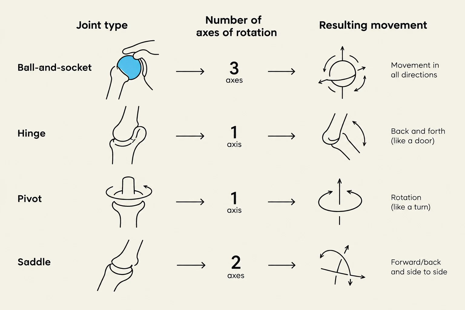
Diarthrodial joints are further classified by their specific geometry and the number of axes (degrees of freedom) they allow:
Joint Type
Description
Axes/Degrees of Freedom
Example
Gliding (Plane)
Nearly flat surfaces; non-axial gliding
Non-axial
Intercarpal joints
Hinge (Ginglymus)
Convex surface fits into concave surface
Uniaxial (1)
Humeroulnar joint
Pivot (Trochoid)
Rotation around a single axis
Uniaxial (1)
Atlantoaxial joint
Condyloid (Ovoid)
Ovular convex and concave shapes
Biaxial (2)
Radiocarpal joint
Saddle (Sellar)
Surfaces shaped like a horse's saddle
Biaxial (2)
Carpometacarpal of thumb
Ball & Socket
Reciprocally convex and concave
Triaxial (3)
Hip and Shoulder
This diagram illustrates four types of synovial joints: ball-and-socket, hinge, pivot, and saddle. It shows the joint type, the number of axes of rotation, and the resulting movement for each.
Summary Table: Degrees of Freedom
Axes
Degrees of Freedom
Movement Capability
One
Uniaxial
1
Two
Biaxial
2
Three
Triaxial
3
By integrating the knowledge of bone geometry, soft tissue support, and mechanical positioning, we can predict how a joint will behave under stress and identify why certain joints are more prone to injury than others.
Keep reading for free.
Create an account to finish this reading and make it part of your study routine.