An explanation of hemoglobin's structure, its role in oxygen transport, and factors influencing oxygen affinity, including cooperativity, Bohr's Law, and variations like fetal hemoglobin and sickle cell anemia.
Hemoglobin is one of the most vital transport proteins in the human body, serving as the primary vehicle for moving oxygen from the lungs to the tissues that need it for cellular respiration. To understand how this protein functions so efficiently, we must look closely at its complex quaternary structure and the chemical environment that dictates its ability to grab and release oxygen.
The Architecture of Hemoglobin
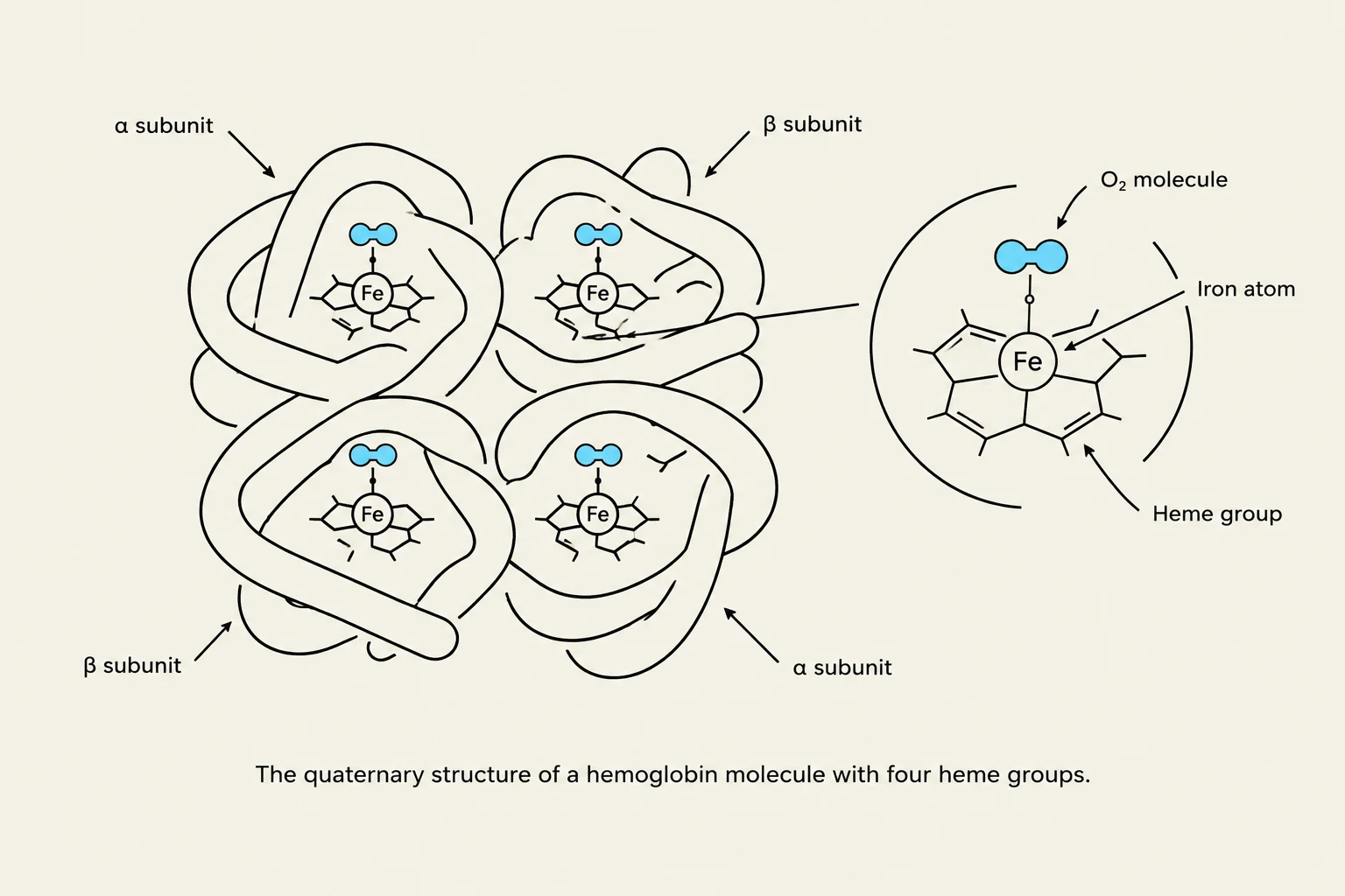
The quaternary structure of a hemoglobin molecule with four heme groups.
Proteins are organized into four levels of structure: primary, secondary, tertiary, and quaternary. While many proteins consist of a single polypeptide chain, hemoglobin is a complex quaternary protein composed of four subunits.
Heme Groups and Iron
Each of these four subunits contains a specialized non-protein component called a heme group. At the very center of each heme group sits an iron atom. This iron atom is the specific site where oxygen (O2) binds. Because there are four subunits, a single hemoglobin molecule can bind up to four O2 molecules. To put this scale into perspective, a single red blood cell typically contains approximately 200 million hemoglobin molecules.
Cooperativity: The Power of Binding
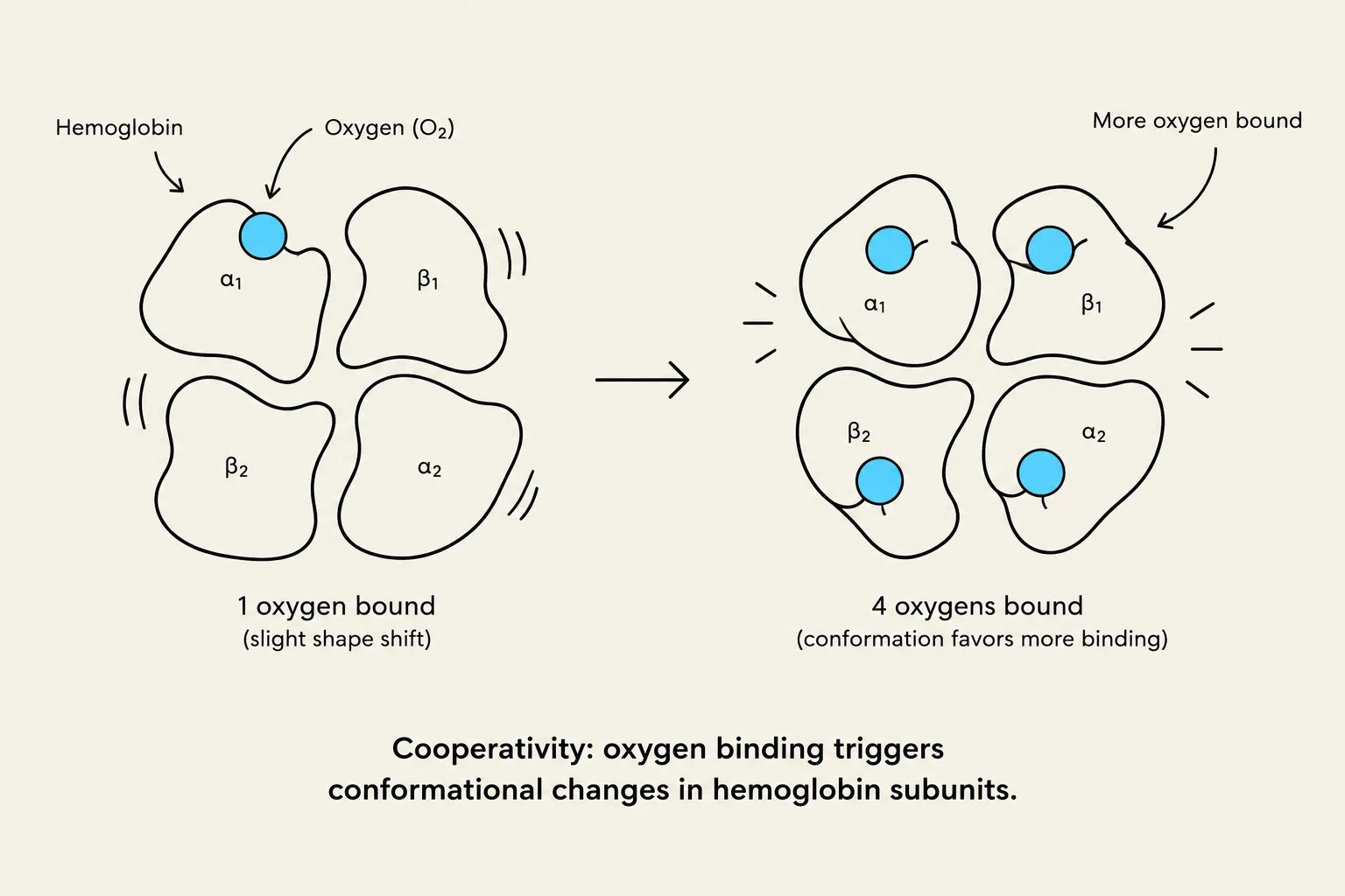
This diagram illustrates the concept of cooperativity in hemoglobin, showing how the binding of oxygen to one subunit can trigger conformational changes that favor the binding of more oxygen to other subunits.
Hemoglobin does not bind oxygen in a simple, linear fashion. Instead, it exhibits a phenomenon known as cooperativity. When the first oxygen molecule binds to one of the heme sites, it causes a conformational change in the protein structure that increases the affinity (the "hunger" or attraction) for oxygen at the remaining three sites. This ensures that once hemoglobin starts picking up oxygen in the lungs, it quickly becomes fully saturated.
Oxygen Transport and Myoglobin
The efficiency of oxygen delivery is driven by the partial pressure of oxygen (PO2).
In the Lungs: The PO2 is high (≈100 mmHg), which encourages hemoglobin to become fully loaded with oxygen.
In the Tissues: The PO2 is much lower (≈40 mmHg), which causes hemoglobin's affinity for oxygen to decrease, allowing the O2 to be released where it is needed most.
The Role of Myoglobin
While hemoglobin transports oxygen through the blood, myoglobin (Mb) serves as an oxygen storage unit within the muscle tissue. Myoglobin has a much higher affinity for oxygen than hemoglobin does, represented by a "left-shifted" dissociation curve. This high affinity creates a chemical gradient: as hemoglobin releases oxygen in the capillaries, the myoglobin in the nearby muscle "pulls" the oxygen away from the hemoglobin and into the muscle cells.
Factors Influencing Oxygen Affinity
The body can fine-tune how much oxygen hemoglobin releases by altering its affinity through various physiological signals. These changes are visualized as shifts in the oxygen-dissociation curve.
Bohr’s Law and Metabolic Demand
During periods of high metabolic activity, such as intense exercise, tissues produce more waste products. This leads to several changes that shift the hemoglobin curve to the right, meaning affinity decreases and oxygen unloading increases:
Bohr’s Law (pH and $H^+$): An increase in hydrogen ion concentration (which results in a low pH) decreases hemoglobin's affinity for O2, promoting more efficient unloading in the tissues.
Carbon Dioxide (CO2): Higher levels of CO2 also shift the curve to the right.
Temperature: Increased body temperature (often seen during exercise) shifts the curve to the right.
Adaptation to Altitude
When humans move to high altitudes where oxygen is scarce, the body compensates by increasing the production of 2,3-DPG. This molecule shifts the curve to the right, facilitating the release of oxygen to the tissues to compensate for the lower environmental oxygen levels.
Exam Tip: Remember that a Right Shift means "Release." If the curve shifts right (due to high $H^+$, high CO2, high Temp, or DPG), hemoglobin's affinity for oxygen decreases, making it easier for oxygen to leave the protein and enter the tissues.
Clinical Variations: Fetal Hemoglobin and Sickle Cell Anemia
Small changes in the amino acid sequence of hemoglobin can lead to massive differences in function or even life-threatening diseases.
Fetal Hemoglobin
In utero, the fetus must extract oxygen from the mother's blood. To do this, fetal hemoglobin has evolved to have a higher affinity for O2 than adult hemoglobin. This higher affinity allows the fetal red blood cells to effectively "pull" oxygen away from the maternal red blood cells.
Sickle Cell Anemia: A Genetic Mutation
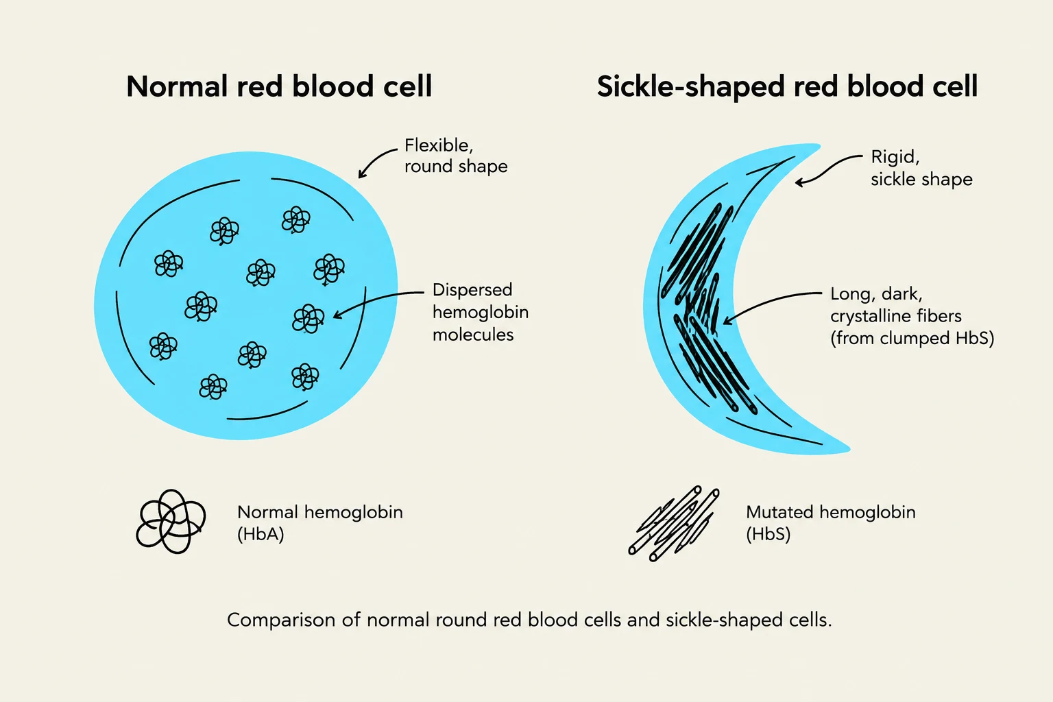
Comparison of normal round red blood cells and sickle-shaped cells, illustrating the difference in hemoglobin structure and cell morphology.
Sickle Cell Anemia is a classic example of how a single genetic mutation can disrupt protein function.
In this condition, a single amino acid substitution occurs in the primary structure of the hemoglobin molecule: the amino acid glutamate (an acidic amino acid) is replaced by valine (a nonpolar amino acid).
This substitution has devastating consequences:
Structural Change: The mutated hemoglobin molecules (Hemoglobin S) interact with one another, crystallizing into long fibers.
Cellular Impact: These fibers distort the red blood cell into a "sickle" shape, which is weak and does not flow well through narrow capillaries.
Health Consequences: The spleen often removes these damaged cells, leading to a lower red blood cell count. However, interestingly, individuals with this trait show an increased resistance to malaria.
Feature
Normal Hemoglobin (HbA)
Sickle Cell Hemoglobin (HbS)
Amino Acid
Glutamate (Glu)
Valine (Val)
RBC Shape
Round, flexible
Sickle-shaped, rigid
Oxygen Capacity
Normal
Greatly reduced
Flow in Capillaries
Smooth
Blocked/Difficult
Keep reading for free.
Create an account to finish this reading and make it part of your study routine.