Foundations of Pathophysiology: From Cells to Disease
An introduction to pathophysiology, covering core terminology, homeostasis, cellular energy metabolism, tissue types, and cellular responses to stress, adaptation, and injury.
Pathophysiology is the study of the mechanisms and processes that alter the natural function of biological systems, ultimately leading to disease. To understand how a person moves from health to illness, one must look beyond symptoms and examine the underlying disruptions in cellular structure, energy production, and the body's ability to maintain internal stability.
Core Terminology of Disease
Before diving into cellular mechanics, it is essential to master the language used to describe how diseases begin and progress.
Etiology: The origin or cause of a disease or abnormal condition.
Pathogenesis: The progression of the disease, tracing the journey from its initial etiology to its clinical manifestations.
Pathophysiology: The specific processes or mechanisms that alter biological functions, resulting in disease.
When investigating a cause, clinicians may categorize it based on its source. If a disease has no known or identifiable cause, it is termed idiopathic. Conversely, if a condition is caused by medical intervention, it is described as iatrogenic.
Homeostasis and the Cycle of Disorder
At the heart of all biological health is homeostasis, a self-regulating process that allows biological systems to maintain a steady state of internal balance. When this balance is disrupted, disease occurs.
Disruptions often manifest through a cycle involving structural disorders (physical changes in the anatomy) and functional disorders (changes in how an organ or system operates).
Clinical Example: The Hypertension Cycle
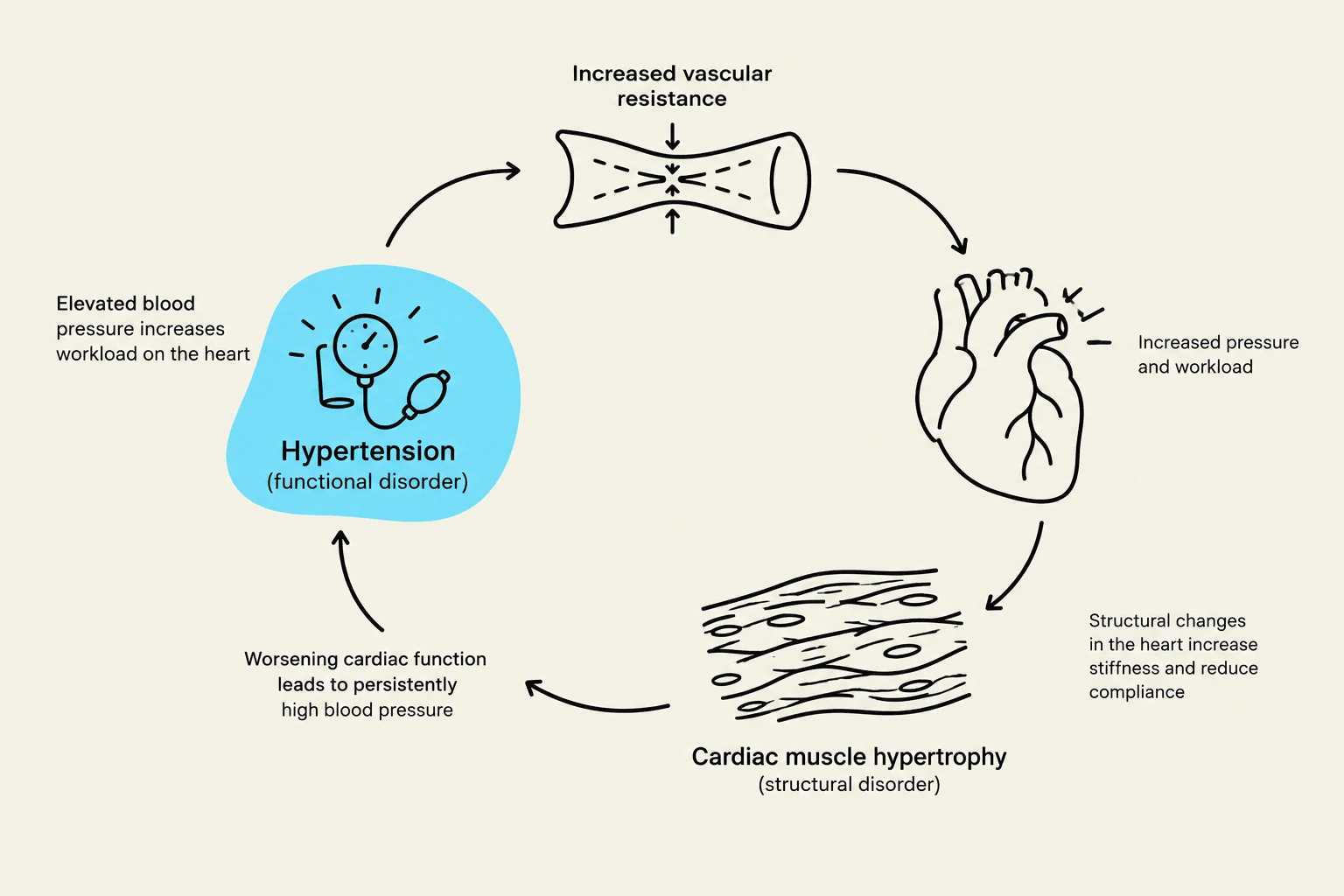
Chronic essential hypertension is a functional disorder where blood pressure remains abnormally high. To cope with this pressure, the heart must contract with more force, leading to a thickening of the heart muscle—a structural disorder. This thickened wall eventually reduces the volume of the ventricle, creating a feedback loop that can lead to congestive heart failure.
This diagram illustrates the cyclical relationship between hypertension, increased vascular resistance, cardiac muscle hypertrophy, and worsening cardiac function.
Cellular Energy and Metabolism
Cells are the fundamental units of life, and their survival depends heavily on their ability to produce energy in the form of Adenosine Triphosphate (ATP). The method of production depends on the availability of oxygen.
Aerobic vs. Anaerobic Metabolism
Most cells utilize aerobic metabolism to produce the vast majority of their ATP. This process occurs in the mitochondria and requires oxygen to act as the final electron acceptor in the Electron Transport Chain (ETC).
If oxygen is unavailable (hypoxia), cells must switch to anaerobic metabolism. While this allows for continued energy production via glycolysis in the cytoplasm, it is far less efficient, producing only a net of 2 ATP per glucose molecule compared to the massive yield of aerobic respiration. In humans, anaerobic metabolism results in the production of lactic acid (lactate).
Clinical Connection: Thiamine Deficiency
Thiamine (Vitamin B1) is a critical cofactor for converting pyruvate into Acetyl CoA, which allows the Krebs cycle and ETC to function. In chronic alcoholics, thiamine absorption is inhibited, preventing the production of ATP. This leads to Wernicke-Korsakoff Syndrome, where metabolically active organs like the brain fail due to energy starvation.
The Building Blocks: Primary Tissue Types
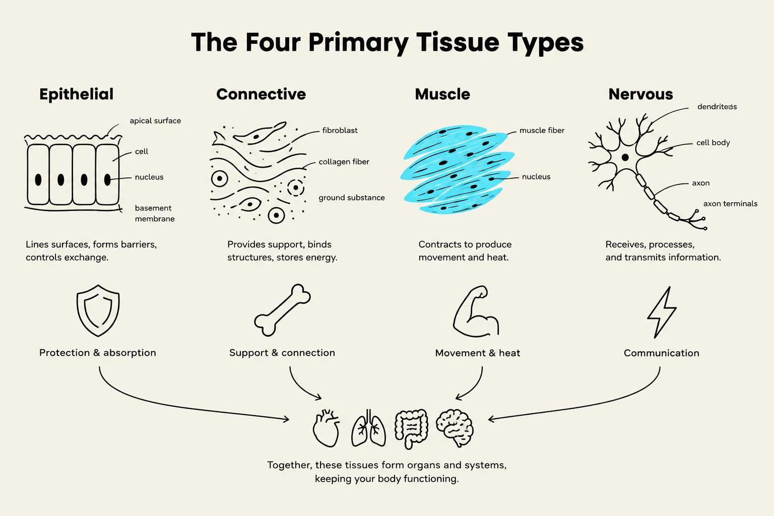
The human body is organized into four primary tissue types, each with a structure specifically designed to support its function:
Epithelial Tissue: Composed of tightly connected cells that cover surfaces, secrete substances, or absorb nutrients. For example, simple squamous epithelium in the alveoli allows for rapid gas exchange.
Connective Tissue: Provides support, protection, and transport. It consists of cells within an extracellular matrix. Examples include bone, cartilage, blood, and adipose tissue.
Muscle Tissue: Specialized for movement. It is divided into skeletal (voluntary), cardiac (involuntary heart muscle), and smooth (involuntary movement in hollow organs).
Nervous Tissue: Composed of neurons (which transmit signals) and neuroglia (which support and protect neurons), facilitating communication and control.
The four primary tissue types in the human body are epithelial, connective, muscle, and nervous tissue. Each type has a distinct structure and function, contributing to the overall organization and operation of organs and systems.
Cellular Response to Stress: Adaptation and Injury
When cells encounter stressors—such as hypoxia, physical trauma, or toxins—they attempt to respond to maintain homeostasis.
Cellular Adaptation
If the stress is chronic but not immediately lethal, cells undergo adaptation to survive:
Atrophy: A decrease in cell size.
Hypertrophy: An increase in cell size.
Hyperplasia: An increase in the number of cells.
Metaplasia: The replacement of one differentiated cell type with another (e.g., respiratory cells changing due to smoking).
Dysplasia: Abnormal changes in the size, shape, and organization of cells. Note that dysplasia is not a true adaptation and can be pre-neoplastic (precancerous).
Reversible vs. Irreversible Injury
If the stressor is removed quickly, the cell may undergo reversible injury, such as cellular swelling (caused by sodium influx) or steatosis (fatty change).
However, if the stress is too severe or prolonged, the cell reaches a point of irreversible injury, leading to cell death. There are two primary pathways for cell death:
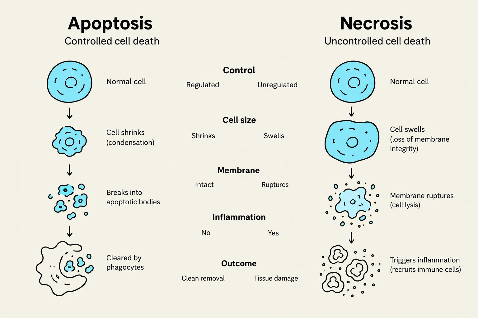
Apoptosis: Programmed, controlled cell death. This is often a physiologic process where the cell shrinks and fragments without causing inflammation.
Necrosis: Uncontrolled cell death resulting from severe injury. The cell membrane ruptures, releasing contents that trigger inflammation. Common patterns include coagulative necrosis (often from ischemia/infarction) and liquefactive necrosis (often seen in brain infarcts or abscesses).
This diagram illustrates the key differences between apoptosis (controlled cell death) and necrosis (uncontrolled cell death), detailing processes such as cell shrinkage, membrane integrity, inflammation, and the final outcome.
Exam Tip: Hypertrophy vs. Hyperplasia
Remember: Hypertrophy is about the size of the individual cells (e.g., a weightlifter's muscles), whereas hyperplasia is about the number of cells (e.g., during lactation).
Keep reading for free.
Create an account to finish this reading and make it part of your study routine.