


In the study of biology and medicine, understanding the microscopic architecture of the body is essential. This field of study is known as histology. Rather than looking at individual cells in isolation, histology examines tissues—groupings of similar cells that work together to perform a common or related function.
The central theme of histology is the relationship between structure and function. The specific arrangement, shape, and density of cells are not random; they are optimized to meet the physiological demands of the organ they compose. When these tissues function correctly, they maintain homeostasis, the self-regulating process by which biological systems maintain a steady state of internal balance. If the ability to manage cellular stress or injury is compromised, the resulting disruption can lead to disease.
Epithelial Tissue: The Body's Coverings and Secretors
Epithelial tissues serve as the primary interface between the body and its environment. These tissues are characterized by tightly connected cells that are avascular, meaning they lack their own blood supply. Because they are often exposed to friction or environmental changes, they possess high regenerative capabilities.
Epithelial tissues are classified based on two main criteria: the number of cell layers and the shape of the cells.
Classification by Layers
- Simple: A single layer of cells, ideal for absorption or rapid diffusion.
- Stratified: Multiple layers of cells, providing protection against abrasion.
- Pseudostratified: A single layer of cells that appears to be layered because the nuclei are at different levels.
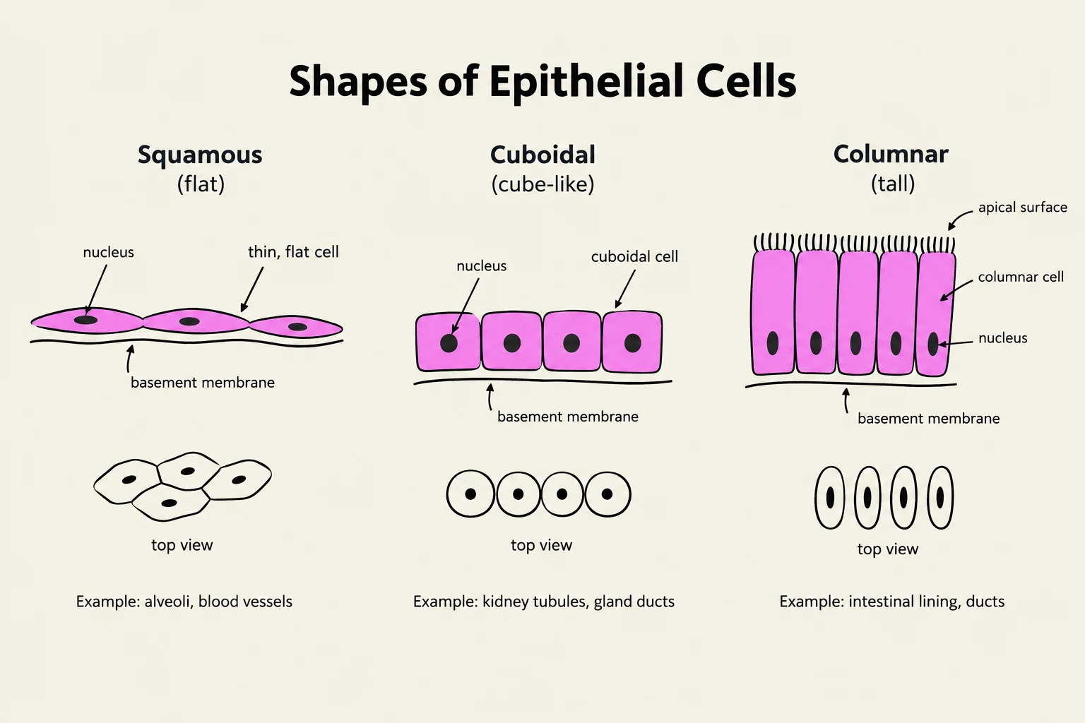
Classification by Shape
- Squamous: Flat, scale-like cells.
- Cuboidal: Cube-shaped cells.
- Columnar: Tall, column-like cells.
Exam Tip: When analyzing a tissue slide, always look for the "free surface" (apical surface). If there is only one layer of cells touching that surface, it is a "simple" epithelium. If you see many layers, it is "stratified".

Keep reading for free.
Create an account to finish this reading and make it part of your study routine.
Continue reading freeAlready have an account? Log in
Updated