


The human skeleton is not a single, rigid structure; rather, it is a complex assembly of bones held together by various types of connective tissues. These connections, known as joints, serve the dual purpose of preventing bones from dislocating and protecting them from breaking one another. Because different parts of the body require different levels of stability and mobility, joints are classified primarily by their degree of movement.
Understanding joint classification is essential for grasping how the body manages mechanical loads, such as compression, tension, and shear, and how it maintains stability during movement.
Synarthrosis: Immovable Joints
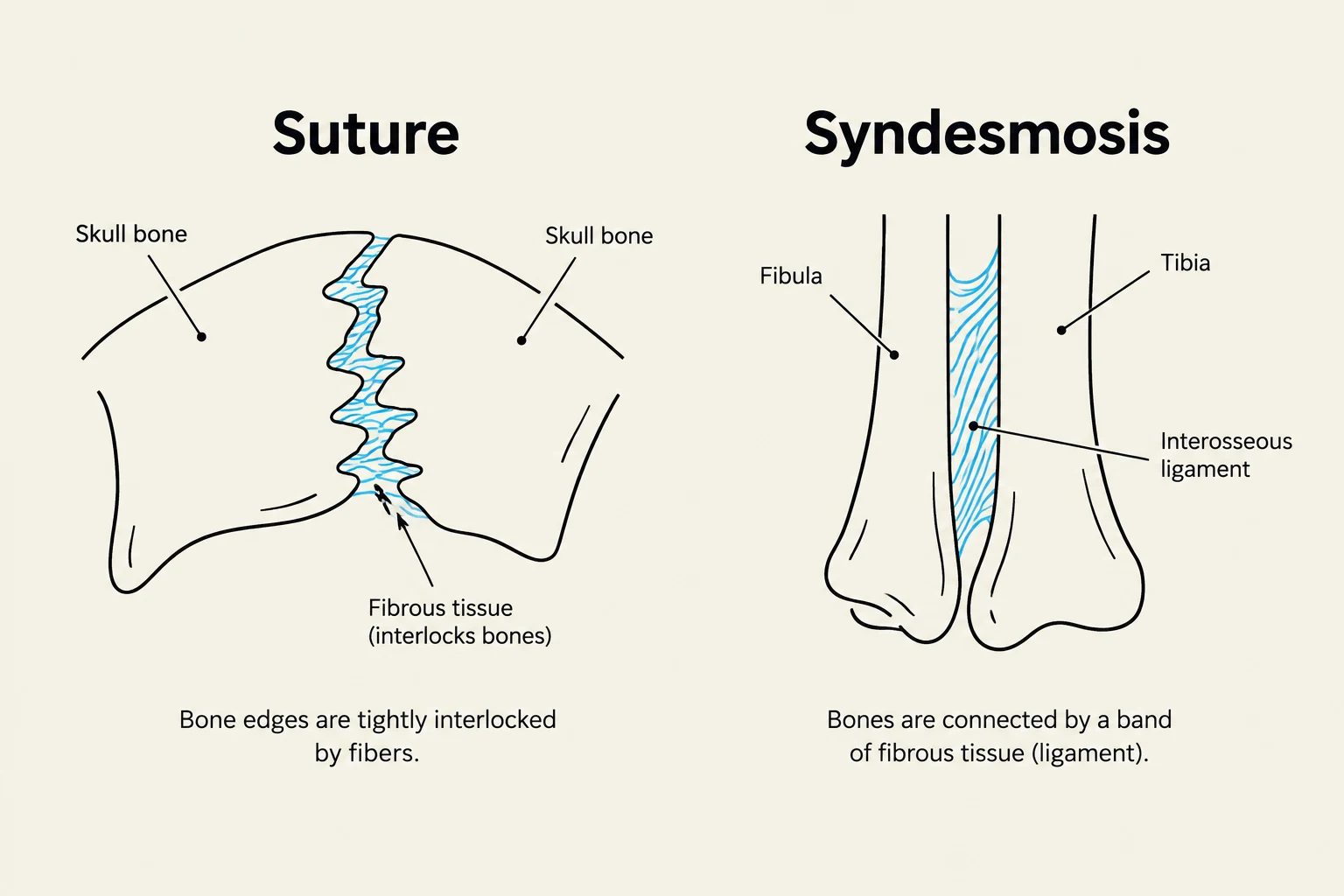
The first category of classification is the Synarthrosis (from the Greek syn, meaning together, and arthron, meaning joint). These are immovable joints designed for maximum stability and the attenuation of shock. Most synarthrodial joints are categorized as fibrous joints, meaning the bones are held together by dense fibrous connective tissue.
There are two primary subcategories of synarthrosis:
- Sutures: These are irregular grooves found on sheets of bone that are tightly connected by fibers continuous with the periosteum. In humans, sutures are found exclusively in the skull. As an individual reaches adulthood, these joints may undergo ossification, effectively turning the connection into solid bone.
- Syndesmosis: These joints are "held by bands" of dense fibrous tissue. While they allow for very limited movement, their primary role is connection. A classic example is the interosseous membrane, which connects the radius and ulna in the forearm, or the tibia and fibula in the lower leg.
Keep reading for free.
Create an account to finish this reading and make it part of your study routine.
Continue reading freeAlready have an account? Log in
Updated