Cardiology: Heart Failure, Cardiomyopathy, and Shock
An overview of cardiology, covering cardiac hemodynamics, heart failure, cardiomyopathy, peripheral vascular disease, and circulatory shock. Includes explanations of stroke volume, ejection fraction, systolic vs. diastolic dysfunction, and types of shock.
Cardiology encompasses a wide range of complex conditions that affect the heart's ability to pump blood and maintain systemic perfusion. Understanding these conditions requires a firm grasp of cardiac hemodynamics—specifically how the heart manages volume and pressure—and how failures in these mechanisms lead to life-threatening states like congestive heart failure and circulatory shock.
Cardiac Hemodynamics: Measuring Heart Function
To understand heart failure, one must first understand how we quantify the heart's efficiency. The heart operates by filling with blood and then contracting to eject that blood into the systemic circulation.
Stroke Volume and Ejection Fraction
The amount of blood ejected from the left ventricle during a single contraction is known as the Stroke Volume (SV). This is determined by the difference between the volume of blood in the heart before it beats and the volume remaining after it beats:
SV=EDV−ESV
Where:
End-Diastolic Volume (EDV): The volume of blood in the ventricle at the end of its filling phase (diastole).
End-Systolic Volume (ESV): The volume of blood remaining in the ventricle after contraction (systole).
The most critical clinical metric for assessing cardiac pump function is the Ejection Fraction (EF). This represents the percentage of the total end-diastolic volume that is successfully pumped out with each beat.
EF=EDVSV
Worked Example: Calculating EF
If a patient has an End-Diastolic Volume (EDV) of 130 mL and an End-Systolic Volume (ESV) of 50 mL:
First, find the Stroke Volume: 130−50=80 mL.
Then, calculate the EF: 13080≈61.5%.
A normal EF is typically around 55–70%. A low EF indicates systolic dysfunction.
Congestive Heart Failure (CHF)
Comparison of Systolic and Diastolic Dysfunction. Systolic dysfunction is characterized by a dilated ventricle with a thin wall that fails to squeeze, resulting in less blood ejected. Diastolic dysfunction involves a thick, muscular wall that is stiff, leading to a small chamber size during filling, despite a normal ejection fraction.
Congestive Heart Failure occurs when the heart's pumping ability is decreased, making it unable to meet the metabolic demands of the body. Heart failure is often categorized by which side of the heart is failing:
Left-Sided Heart Failure: When the left ventricle fails, blood backs up into the pulmonary circulation (the lungs). This leads to Pulmonary Edema, a buildup of fluid in the lung tissue that makes breathing difficult.
Right-Sided Heart Failure: When the right ventricle fails, blood backs up into the systemic venous circulation. This results in Pitting Edema (swelling in the extremities) and potentially ascites.
Cor Pulmonale: This is a specific type of right-sided heart failure that is caused by a primary respiratory disorder, such as COPD or pulmonary fibrosis, rather than a primary heart disease.
Systolic vs. Diastolic Dysfunction
Heart failure can also be classified by the specific mechanical failure occurring in the myocardium:
Systolic Dysfunction: The heart muscle is too weak to contract effectively. This results in a low Ejection Fraction and is often associated with an S3 heart sound. The most common cause is Dilated Cardiomyopathy.
Diastolic Dysfunction: The heart muscle is too stiff to relax and fill properly. In these cases, the Ejection Fraction remains normal, but the volume of blood entering the heart is limited. This is often associated with an S4 heart sound and is commonly caused by Hypertrophic Cardiomyopathy.
Cardiomyopathy
Cardiomyopathy refers to diseases of the myocardium (the heart muscle) that impair its function. There are three primary types:
Type
Description
Dysfunction Type
Common Causes
Dilated
All four chambers are enlarged/dilated
Systolic (Low EF)
Alcoholism, Ischemic heart disease, Idiopathic
Hypertrophic
Abnormally thickened walls (LV and Septal)
Diastolic (Normal EF)
Hypertension, Autosomal dominant genetics
Restrictive
Abnormally stiff walls due to fibrosis
Diastolic (Normal EF)
Amyloidosis, Hemochromatosis
Peripheral Vascular Disease (PVD)
While heart failure affects the pump, Peripheral Artery Disease (PAD) affects the delivery system to the limbs. PAD is characterized by atherosclerosis in the arteries that supply the legs.
A hallmark symptom of PAD is Intermittent Claudication, which is muscle pain or weakness in the legs that occurs during walking because the narrowed arteries cannot provide enough oxygenated blood to meet the increased demand of exercise.
Clinical Testing: The Ankle-Brachial Index (ABI)
To diagnose PVD, clinicians use the Ankle-Brachial Index (ABI). This is a ratio of the systolic blood pressure at the ankle to the systolic blood pressure in the arm:
ABI=Systolic BParmSystolic BPankle
Normal:≈1.0
PVD Indicated:<0.9 (This means the blood pressure in the arm is higher than in the ankle, suggesting a blockage in the leg).
Circulatory Shock
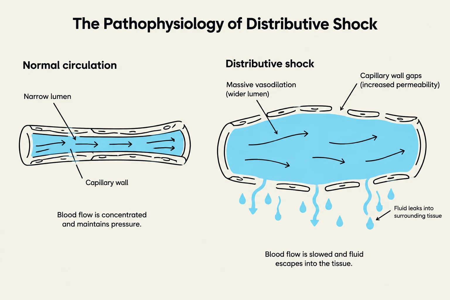
This diagram illustrates the difference between normal circulation and distributive shock, highlighting how vasodilation and increased capillary permeability lead to fluid leakage and slowed blood flow in distributive shock.
Shock is a life-threatening medical emergency defined by inadequate tissue perfusion, leading to tissue hypoxia (low oxygen) and cellular injury. Regardless of the cause, common signs include hypotension (low BP), tachycardia (high HR), cold/clammy skin, and altered mental status.
Types of Shock
Shock is categorized by its underlying physiological mechanism:
Hypovolemic Shock: Caused by a loss of blood volume, such as from trauma, severe burns, or dehydration.
Cardiogenic Shock: Caused by the heart's inability to pump effectively, often due to heart failure or a recent myocardial infarction.
Distributive Shock: This category includes:
Anaphylactic Shock: A severe allergic reaction where massive histamine release causes widespread vasodilation and increased capillary permeability.
Septic Shock: A systemic infection where bacterial toxins cause massive inflammation, leading to widespread vasodilation and fluid leaking from vessels.
The Progression of Shock
The body's response to shock occurs in two main stages:
Compensated Shock: The body attempts to maintain homeostasis. A drop in blood pressure triggers the baroreflex and the Renin-Angiotensin-Aldosterone System (RAAS). This results in increased heart rate (tachycardia) and vasoconstriction to raise blood pressure and cardiac output.
Decompensated Shock: The compensatory mechanisms fail. Positive feedback loops begin to drive the patient further into crisis—for example, low cardiac output leads to myocardial ischemia, which further decreases cardiac output.
Keep reading for free.
Create an account to finish this reading and make it part of your study routine.