Biology is often defined by the study of life, but some of the most influential entities in our world exist on the very edge of that definition. Viruses, though not technically living organisms, interact with living cells in complex ways that drive evolution and disease. On the other side of this biological spectrum are eukaryotic cells—highly organized, complex units of life that use internal compartmentalization to perform the sophisticated tasks necessary for survival. Understanding the relationship between these microscopic invaders and the intricate architecture of the eukaryotic cell is fundamental to modern biology.
The Nature of Viruses
Viruses are microscopic entities that differ significantly from the living organisms they infect. Unlike cells, they lack the machinery for independent metabolism and reproduction. Instead, they rely on the hijacked resources of a host cell to propagate.
Size, Shape, and Genetic Material
Viruses are incredibly small, generally much smaller than the cells they inhabit. They exhibit a wide variety of shapes and sizes, ranging from simple geometric shells to more complex structures. A defining characteristic of a virus is its genetic material; depending on the type of virus, the genome may consist of either DNA or RNA. This distinction is crucial because it dictates how the virus will interact with the host's molecular machinery to replicate.
Enveloped vs. Non-Enveloped Viruses
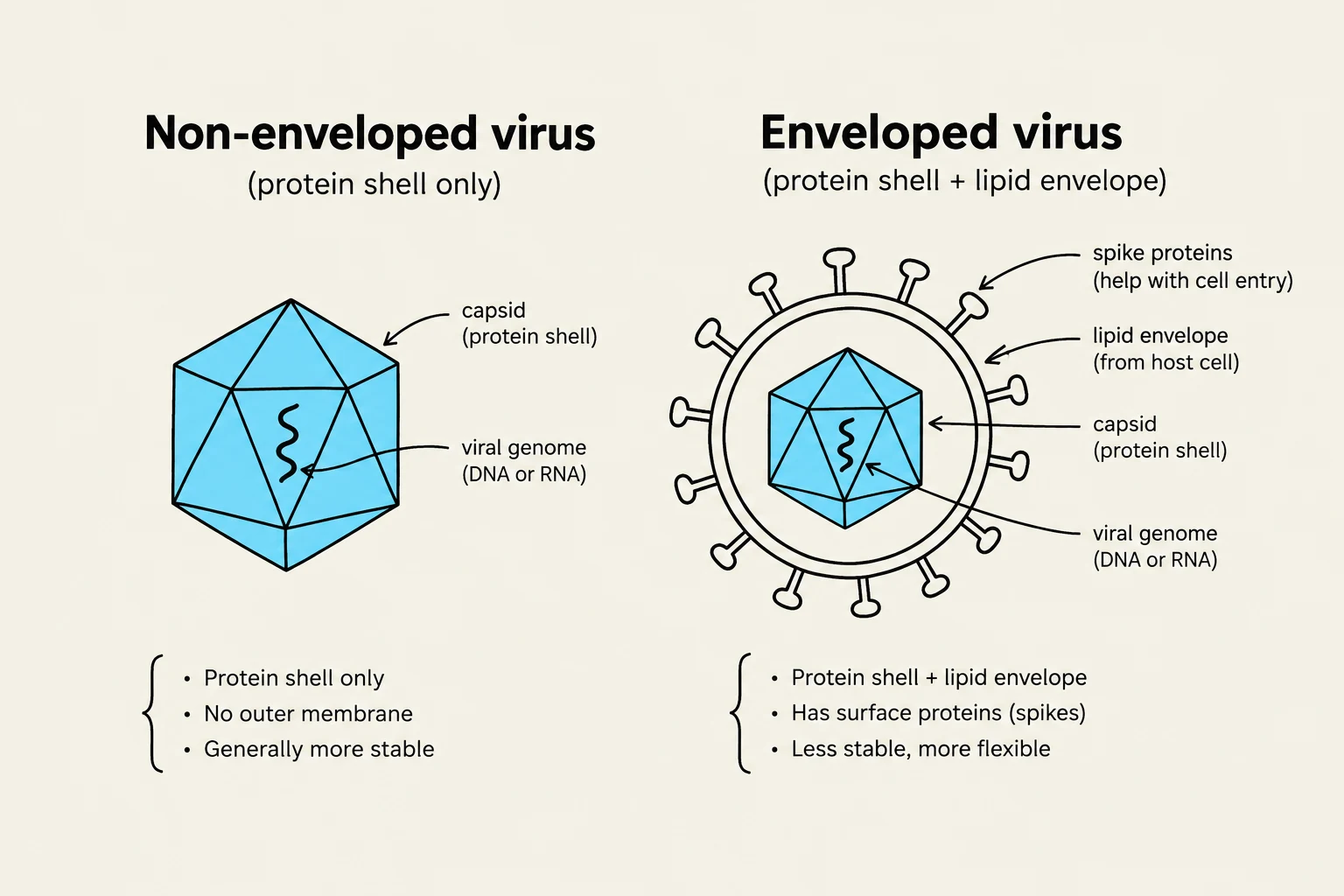
Viruses are further classified by their outer structure. Some viruses possess a lipid membrane derived from the host cell, which classifies them as enveloped viruses. A well-known example is HIV, which utilizes its envelope to interact with the host cell.
Exam Tip: Pay close attention to how viruses enter cells. While some viruses enter via endocytosis, enveloped viruses often enter through fusion, where the viral membrane merges directly with the host cell membrane.
Diagram illustrating the structural differences between non-enveloped and enveloped viruses, detailing components like the capsid, viral genome, and lipid envelope.
The Viral Life Cycle: Replication and Exit
The life cycle of a virus is a systematic process of hijacking, copying, and escaping. This cycle generally follows three main stages: entering the host cell, reproducing the viral components, and leaving the cell to infect new hosts.
Viral Propagation and the Lytic Cycle
Once inside, the virus takes control of the cell's metabolic pathways to produce new viral progeny. In many cases, this leads to the lytic cycle, where the host cell is essentially turned into a virus factory. The goal of this stage is rapid propagation—ensuring that as many copies of the viral genome and proteins are made as possible.
Mechanisms of Exit: Budding and Bursting
To complete the cycle, the newly formed viruses must exit the host cell. They typically use one of two primary mechanisms:
Budding: The virus exits by wrapping itself in a portion of the host's own membrane. This is common for enveloped viruses, as it allows them to acquire their lipid coating during the exit process.
Bursting: The virus causes the host cell to rupture (lyse), releasing all the viral progeny at once. This process is usually fatal to the host cell.
Impact on Humans
The impact of viruses on human biology is profound. They can infect a vast array of different tissues and are the primary drivers of modern epidemics and pandemics. Furthermore, viruses play a surprising role in our genetic history; they can contribute to human evolution by integrating viral DNA directly into the host genome.
Eukaryotic Cell Compartmentalization
While viruses are simple and "minimalist," eukaryotic cells—which include both animal and plant cells—are defined by their immense complexity. The hallmark of a eukaryotic cell is the presence of various organelles.
The Advantage of Compartments
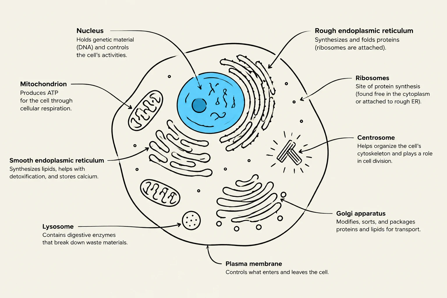
Organelles are membrane-bound compartments that divide the cell into distinct functional zones. This organization provides several critical evolutionary advantages:
Separation of Incompatible Reactions: Compartmentalization allows the cell to perform conflicting chemical reactions simultaneously without them interfering with one another.
Specialized Environments: Different organelles can maintain specific conditions, such as unique pH levels, that are optimized for specific biochemical processes.
Concentration of Substrates: By confining specific enzymes and substrates within a small space, the cell increases the efficiency of metabolic pathways through higher local concentrations.
Size Management: Compartmentalization helps manage the challenges of diffusion and transport, allowing eukaryotic cells to achieve a much larger size than prokaryotic cells.
Diagram of an animal cell with its organelles labeled, including the nucleus, mitochondria, endoplasmic reticulum, ribosomes, centrosome, Golgi apparatus, lysosome, and plasma membrane.
Comparing Animal and Plant Cells
Although both are eukaryotic and share the fundamental principle of compartmentalization, they possess distinct structures. For example, plant cells contain chloroplasts for photosynthesis and a large central vacuole for storage and structural support, whereas animal cells lack these specific features.
Note on Energy: In all eukaryotes, the mitochondria serve as the central hub for aerobic respiration, converting energy from food into a usable form, regardless of whether the cell is part of a plant or an animal.
Summary Table: Viruses vs. Eukaryotic Cells
Feature
Viruses
Eukaryotic Cells
Living Status
Non-living (biological entities)
Living organisms
Genetic Material
DNA or RNA
DNA (contained in nucleus)
Size
Extremely small (nanometers)
Larger (micrometers)
Organization
Simple (capsid/envelope)
Complex (organelles/compartments)
Reproduction
Requires a host cell
Independent reproduction
Keep reading for free.
Create an account to finish this reading and make it part of your study routine.