An overview of viral structure, including helical and icosahedral symmetries, enveloped vs. naked viruses, and the five-step replication cycle: attachment, penetration, synthesis, assembly, and release. It also covers the Baltimore classification, retroviruses, lytic vs. lysogenic cycles, and specialized viral enzymes.
Viruses are obligate intracellular parasites, meaning they are genetic elements that can only multiply within a living host cell. Because they rely entirely on a host for energy, metabolic intermediates, and protein synthesis, they are not considered "living" and do not fit into the traditional tree of life. A complete, infectious virus particle outside of a host cell is known as a virion. While viruses vary wildly in size—from the tiny Poliovirus (~28 nm) to the massive Pandoravirus (>1 μm)—most fall within the range of 0.02 to 0.3 μm.
The Architecture of a Virion
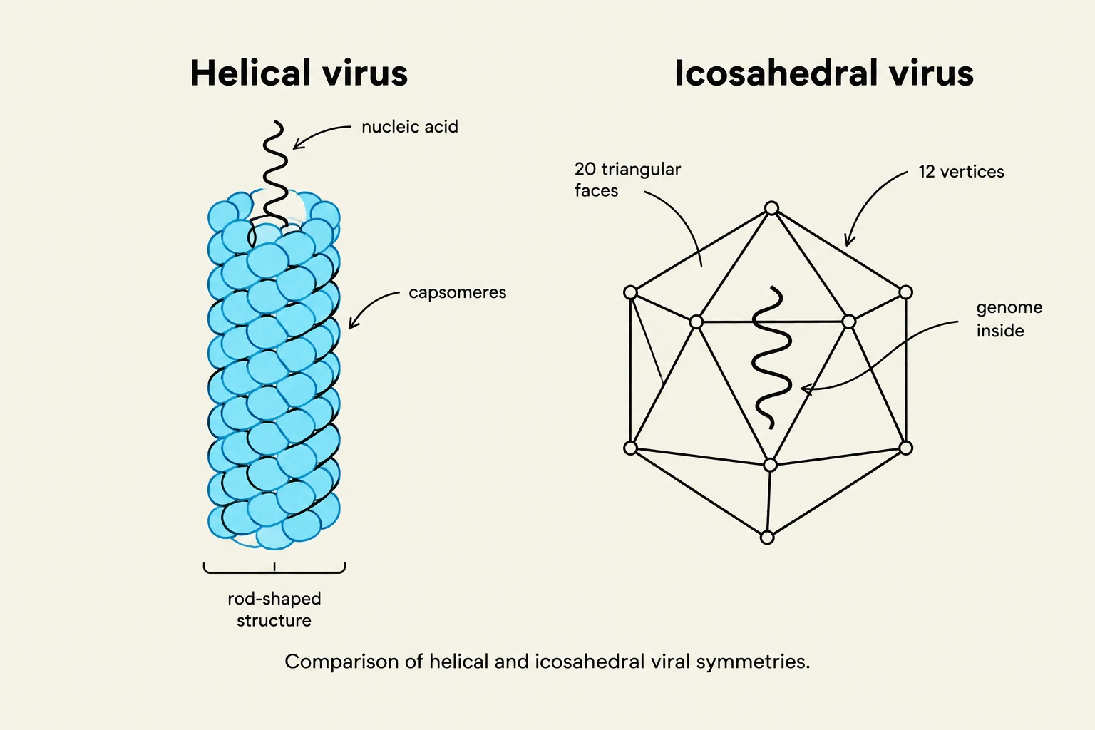
Comparison of helical and icosahedral viral symmetries, illustrating the arrangement of capsomeres and nucleic acid in helical viruses and the icosahedral structure with internal genome.
The fundamental structure of a virus consists of a nucleic acid genome (either DNA or RNA) enclosed within a protein shell called a capsid. The individual protein subunits that make up the capsid are called capsomeres. These subunits are arranged in precise, repetitive patterns that can form through spontaneous self-assembly or with the assistance of host cell protein-folding machinery.
Viruses generally exhibit two primary types of symmetry:
Helical symmetry: A rod-shaped structure where the length is determined by the nucleic acid and the width by the capsomeres (e.g., Tobacco Mosaic Virus).
Icosahedral symmetry: A spherical shape consisting of 20 triangular faces and 12 vertices. This is the most efficient arrangement for a closed shell, requiring the fewest capsomeres.
Some viruses possess highly complex structures. Bacteriophages (viruses that infect bacteria) often feature a "head-plus-tail" morphology, such as the T4 phage, which includes tail fibers and a base plate. Other large viruses, like the Mimivirus or Pandoravirus, utilize specialized portals such as a "stargate" or an apical pore to release their genome.
Enveloped vs. Naked Viruses
Viruses are further categorized by the presence or absence of an outer layer:
Naked viruses: These consist only of the nucleocapsid (nucleic acid + capsid). Most bacterial and plant viruses are naked.
Enveloped viruses: These possess an outer lipoprotein membrane (a phospholipid bilayer derived from the host cell) surrounding the nucleocapsid. Enveloped viruses, such as Influenza or Ebola, use surface proteins to facilitate attachment.
Exam Tip: Enveloped viruses generally exit host cells more easily via budding, but they are less common in plants and bacteria because the rigid cell walls of those organisms make membrane acquisition difficult.
The Five Steps of Viral Replication
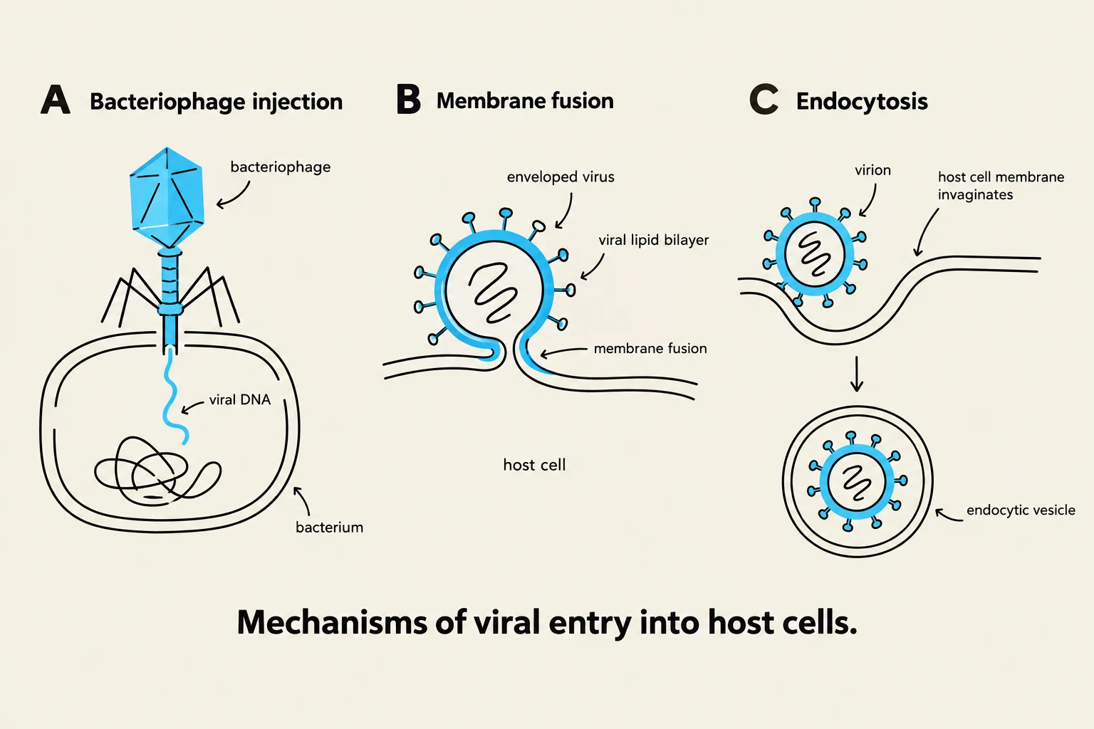
Diagrams illustrating three mechanisms of viral entry into host cells: bacteriophage injection, membrane fusion, and endocytosis.
Regardless of the host, all viral replication follows a fundamental five-step cycle:
Attachment (Adsorption): The virion binds to specific host cell receptors. This is a "lock and key" mechanism where a viral protein or glycoprotein must match a specific protein or carbohydrate on the host surface. This specificity is why viruses often only infect certain tissues.
Penetration (Entry):
In bacteriophages and naked animal viruses, this often occurs via direct penetration, where the genome is injected into the cell. For example, the T4 bacteriophage uses lysozyme to create a hole in the bacterial cell wall and then "shoots" its DNA into the cytoplasm like a syringe.
In enveloped animal viruses, entry occurs via endocytosis (the whole virion is taken in via a vesicle) or membrane fusion (the viral envelope fuses with the host membrane, releasing the capsid into the cell).
Synthesis: The virus hijacks the host's metabolic machinery (enzymes, nucleotides, amino acids, and ATP) to replicate its genome and translate viral proteins.
Assembly (Maturation): Viral parts (capsids and genomes) are chemically attracted to one another and packaged into new virions. In some phages, this involves an energy-linked packaging motor that uses ATP to pump DNA into a "prohead".
Release:
Lysis: The host cell bursts, releasing the new virions. This is common in bacteriophages and naked animal viruses.
Budding: Enveloped viruses push through the host membrane, taking a piece of it with them to form their new envelope. This is a slower process that can lead to persistent infections.
The Baltimore Classification and Specialized Strategies
Because viruses use different types of nucleic acids, they require different strategies to produce mRNA and replicate their genomes. The Baltimore classification system categorizes viruses based on these genomic differences.
Retroviruses and Reverse Transcription
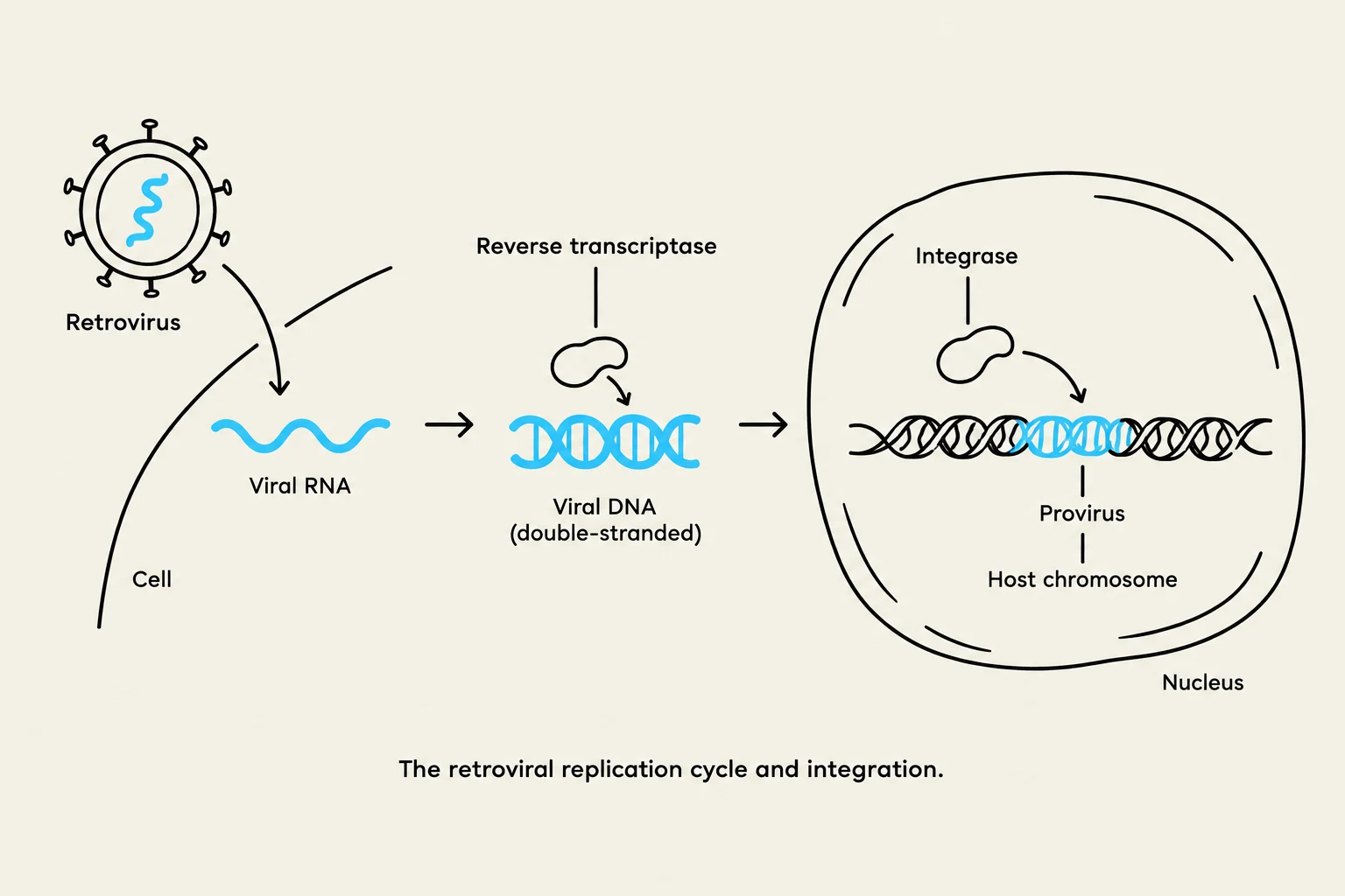
The retroviral replication cycle and integration.
A unique group of viruses, known as retroviruses (e.g., HIV), uses an RNA genome but must convert it into DNA to replicate. They carry a specialized enzyme called reverse transcriptase (an RNA-dependent DNA polymerase).
The process involves:
Reverse Transcription: The viral RNA is used as a template to synthesize complementary DNA (cDNA).
Integration: The enzyme integrase helps the viral DNA insert itself into the host cell's chromosome. Once integrated, the virus is known as a provirus (in animal cells) or a prophage (in bacteria).
Lytic vs. Lysogenic Cycles
In bacteria, viruses can follow two distinct pathways:
Lytic Cycle: The virus immediately undergoes synthesis and assembly, ending in the lysis and death of the host cell.
Lysogenic Cycle: The virus establishes a stable, long-term relationship with the host. The viral genome (prophage) is replicated along with the host's DNA and passed to offspring without killing the host.
Note: A lysogenic infection can be reversed through induction. Environmental stressors (like DNA damage) can cause the viral genome to detach from the host chromosome and re-enter the lytic cycle.
Infection Outcomes in Animal Cells
In eukaryotic hosts, the outcomes of infection can vary:
Virulent Infection: Rapid replication followed by cell lysis.
Latent Infection: The virus exists as a provirus in the host genome, remaining dormant until triggered.
Persistent Infection: The virus exits via slow budding, allowing the cell to remain alive and continue producing virions for a long time.
Transformation: The virus inserts itself into the host genome in a way that disrupts normal cell cycle control (e.g., turning a proto-oncogene into an oncogene), potentially leading to cancer.
Specialized Viral Enzymes
To facilitate these complex cycles, many viruses carry or encode specialized enzymes:
Lysozyme: Used by bacteriophages to degrade the peptidoglycan layer of cell walls.
Neuraminidases: Found in viruses like Influenza to destroy host glycoproteins/glycolipids, allowing new virions to be liberated.
RNA Replicases: RNA-dependent RNA polymerases used by RNA viruses to replicate their genomes.
Reverse Transcriptase: Used by retroviruses to convert RNA into DNA.
Keep reading for free.
Create an account to finish this reading and make it part of your study routine.