Ventilation vs. Oxygenation: Understanding Respiratory Mechanics and Gas Exchange
This content explains the critical distinction between ventilation (the mechanical movement of air) and oxygenation (the transfer of oxygen into the blood), crucial for respiratory care and assessment.
In clinical practice, the terms "ventilation" and "oxygenation" are frequently used interchangeably by students and even some healthcare professionals. However, in the context of respiratory physiology and critical care, they represent two distinct, though deeply interconnected, processes. Confusing the two can lead to significant errors in assessment and treatment—for instance, providing supplemental oxygen to a patient who is actually suffering from inadequate ventilation. To master respiratory care, one must first master the distinction between the mechanical movement of air and the biochemical transfer of gases.
The Mechanics of Ventilation
Ventilation is a mechanical process. It refers to the physical movement of air into and out of the lungs, a process driven by pressure gradients between the atmosphere and the alveoli. This cycle is divided into two phases: inspiration (inhalation) and expiration (exhalation).
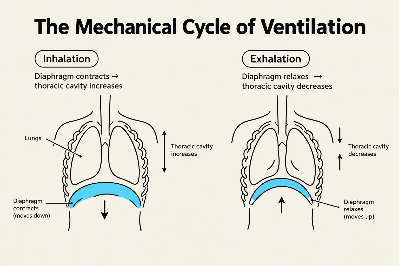
During inspiration, the diaphragm and external intercostal muscles contract, increasing the volume of the thoracic cavity. According to Boyle's Law, as volume increases, pressure decreases. This creates a negative pressure relative to the atmosphere, drawing air into the lungs. During expiration, these muscles relax, the thoracic volume decreases, and the resulting increase in pressure pushes air out of the lungs.
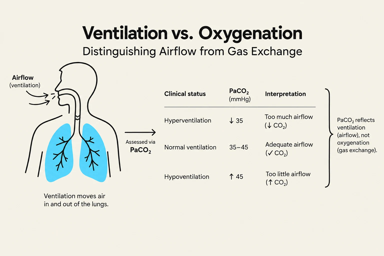
When we discuss "adequate ventilation," we are primarily concerned with the effective exchange of carbon dioxide (CO2). Because CO2 is a byproduct of metabolism that must be "blown off," the adequacy of ventilation is measured by how well a patient can remove CO2 from the bloodstream via the lungs.
Diagram illustrating the mechanical cycle of ventilation, showing the processes of inhalation and exhalation involving the diaphragm and lungs.
Exam Tip: If a question asks about "minute ventilation," it is referring to the total volume of air moved in one minute, calculated as:
Minute Ventilation=Tidal Volume⋅Respiratory Rate
If a patient has a high respiratory rate but very shallow breaths (low tidal volume), their total ventilation may still be inadequate.
The Physiology of Oxygenation
While ventilation is about moving air, oxygenation is about moving oxygen from that air into the blood. Oxygenation occurs at the alveolar-capillary membrane through a process called diffusion. Once air reaches the alveoli, oxygen molecules must cross the thin membrane of the alveoli and the capillary wall to bind with hemoglobin in the red blood cells.
Oxygenation can be compromised even if ventilation is perfect. For example, if a patient is breathing deeply and frequently (excellent ventilation) but has fluid in their lungs (pulmonary edema), the oxygen molecules cannot physically cross the fluid barrier to reach the blood. In this scenario, the patient is well-ventilated but poorly oxygenated.
Diagram illustrating the diffusion of oxygen molecules from an alveolus into a capillary, where they bind to hemoglobin in red blood cells.
Key Clinical Indicators: PaCO2 vs. PaO2
To differentiate these two processes in a clinical setting, we look at Arterial Blood Gas (ABG) results. This is the most common area where students trip up during exams.
1. Carbon Dioxide (PaCO2) as a Marker of Ventilation
The partial pressure of arterial carbon dioxide (PaCO2) is the gold standard for assessing ventilation.
Hypercapnia (High PaCO2): Indicates hypoventilation. The patient is not moving enough air to clear the CO2 produced by the body.
Hypocapnia (Low PaCO2): Indicates hyperventilation. The patient is moving more air than is necessary to meet metabolic demands, "blowing off" too much CO2.
2. Oxygen (PaO2 and SaO2) as a Marker of Oxygenation
The partial pressure of arterial oxygen (PaO2) and oxygen saturation (SaO2) are the markers for oxygenation.
Hypoxemia (Low PaO2): Indicates inadequate oxygenation. This could be due to poor gas exchange at the membrane, low inspired oxygen concentration (FiO2), or ventilation-perfusion (V/Q) mismatch.
This diagram illustrates the difference between ventilation (airflow) and oxygenation (gas exchange), showing how PaCO2 levels relate to airflow in different clinical statuses.
Edge Cases: When the Two Diverge
Understanding the divergence between these two concepts is critical for deciding on an intervention. Consider these two clinical scenarios:
Scenario A: The Opioid Overdose
A patient presents with a very low respiratory rate (4 breaths per minute) and shallow breathing.
Assessment: The patient is hypoventilating. Because they aren't moving enough air, CO2 will build up (High PaCO2). Consequently, oxygenation will eventually fail (Low PaO2) because the "pump" isn't working.
Primary Problem: Ventilation.
Intervention: Respiratory stimulation (e.g., Naloxone) or mechanical ventilation to restore the mechanical movement of air.
Scenario B: The Pneumonia Patient
A patient is breathing rapidly and deeply (28 breaths per minute), but their skin is cyanotic (blue) and they are struggling to stay awake.
Assessment: The patient is ventilating well (they are moving plenty of air, so PaCO2 might actually be low due to hyperventilation), but they are poorly oxygenated (the oxygen cannot cross the inflamed, fluid-filled alveoli).
Primary Problem: Oxygenation.
Intervention: Supplemental oxygen (FiO2) or Positive End-Expiratory Pressure (PEEP) to help keep alveoli open and improve gas exchange.
By separating the "pump" (ventilation) from the "exchange" (oxygenation), clinicians can more accurately diagnose the underlying cause of respiratory distress and apply the correct life-saving interventions.
Keep reading for free.
Create an account to finish this reading and make it part of your study routine.