Vascular Physiology and Cardiovascular Pharmacology
An in-depth exploration of vascular smooth muscle, autonomic nervous system, and hormonal cascades in regulating blood pressure and cardiac output, including mechanisms of contraction, relaxation, hypertension types, RAAS, adrenergic pharmacology, and various drug classes.
Understanding the regulation of blood pressure and cardiac output requires a deep dive into the interplay between vascular smooth muscle, the autonomic nervous system, and complex hormonal cascades. From the microscopic movement of calcium ions within a cell to the systemic administration of potent vasopressors, cardiovascular pharmacology is a study of precision and physiological balance.
Vascular Physiology: Tone and Contraction
The fundamental unit of vascular control is the smooth muscle cell within the arterial and venous walls. The "tone" of these vessels—the degree of constriction or dilation—is primarily governed by the intracellular concentration of calcium (Ca2+).
Mechanisms of Contraction and Relaxation
Contraction is typically driven by the IP3/DAG pathway. When a stimulus activates a Gq-coupled receptor (such as the α1-adrenergic receptor), it triggers the release of IP3, which in turn causes Ca2+ to be released from the sarcoplasmic reticulum. This Ca2+ binds to calmodulin, forming a complex that activates myosin light chain kinase (MLCK), leading to cross-bridge cycling and muscle contraction.
Relaxation occurs through several pathways:
Phosphodiesterase Inhibition: Increasing levels of cAMP or cGMP activate phosphatases that dephosphorylate the myosin light chain.
Calcium Sequestration: The SERCA pump actively moves Ca2+ back into the sarcoplasmic reticulum.
Nitric Oxide (NO) Pathway: NO diffuses into smooth muscle and activates guanylyl cyclase, increasing cGMP.
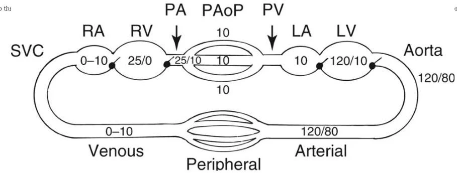
Diagram illustrating blood pressure values in different parts of the circulatory system, including the superior vena cava (SVC), right atrium (RA), right ventricle (RV), pulmonary artery (PA), pulmonary artery occlusion pressure (PAoP), pulmonary vein (PV), left atrium (LA), left ventricle (LV), and aorta.
Hypertension: Primary vs. Secondary
Hypertension (HTN) is a chronic elevation of systemic blood pressure that increases the workload on the heart and damages the vasculature. It is clinically categorized into two types:
Primary (Essential) Hypertension: This accounts for approximately $95%$ of cases and is characterized by a complex interplay of genetic predisposition and environmental factors.
Secondary Hypertension: This is caused by an identifiable underlying condition, such as renal artery stenosis or endocrine disorders, and is often potentially curable.
The Renin-Angiotensin-Aldosterone System (RAAS)
The RAAS is a critical hormonal cascade for long-term blood pressure regulation. The process begins with the release of renin from the juxtaglomerular apparatus in the kidneys. Renin converts angiotensinogen to Angiotensin I, which is then converted to the potent vasoconstrictor Angiotensin II by the Angiotensin-Converting Enzyme (ACE).
Pharmacological Blockade of RAAS
To manage hypertension and heart failure, several classes of drugs target this system:
ACE Inhibitors (e.g., Enalapril): These prevent the formation of Angiotensin II. A common side effect is a dry cough, caused by the accumulation of bradykinin.
Angiotensin II Receptor Blockers (ARBs) (e.g., Losartan): These block the AT1 receptors directly, providing similar benefits to ACE inhibitors without the cough.
Renin Inhibitors (e.g., Aliskiren): These act at the very top of the cascade to prevent the initial conversion of angiotensinogen.
Adrenergic Pharmacology: Alpha and Beta Receptors
The sympathetic nervous system regulates vascular tone through catecholamines (epinephrine, norepinephrine, and dopamine) acting on G-protein coupled receptors.
Adrenergic Receptor Profiles
Receptor
G-Protein
Primary Effect
Clinical Context
α1
Gq
Vasoconstriction (↑Ca2+)
Increases SVR and BP
α2
Gi
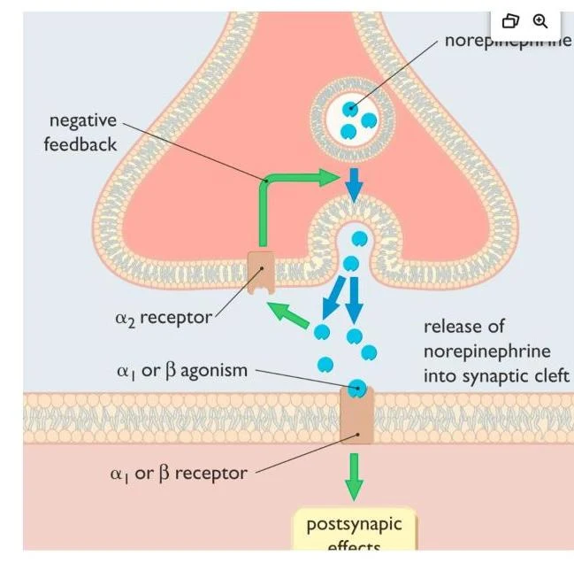
Inhibits NE release
Presynaptic feedback inhibition
β1
Gs
↑ HR, ↑ Contractility
Cardiac stimulation
β2
Gs
Vasodilation, Bronchodilation
Decreases SVR
Diagram illustrating the release of norepinephrine into the synaptic cleft and its interaction with alpha-2 receptors, leading to negative feedback.
Key Vasopressors and Inotropes
Norepinephrine (Levophed): A potent α1 and β1 agonist. It is the first-line treatment for septic shock due to its intense vasoconstriction, which increases MAP.
Epinephrine (Adrenalin): A non-selective agonist (α and β). At low doses, β2 effects predominate (vasodilation); at high doses, α effects predominate (vasoconstriction). It is the drug of choice for anaphylaxis.
Dobutamine: Primarily a β1 agonist used to improve cardiac output in cardiogenic shock without significantly increasing afterload.
Isoproterenol (Isuprel): A pure β1/β2 agonist with no α activity. It acts as a "liquid pacemaker" but can cause significant hypotension due to β2 vasodilation.
Exam Tip: When comparing Epinephrine and Norepinephrine, remember that Norepinephrine is generally more potent at α receptors, while Epinephrine has stronger β1 (inotropic) and β2 (metabolic/bronchodilatory) effects.
Calcium Channel Blockers (CCBs)
CCBs inhibit the influx of calcium through L-type channels, leading to muscle relaxation. They are divided into two main groups:
Dihydropyridines (e.g., Nicardipine, Clevidipine): These are potent arterial vasodilators used primarily for hypertensive emergencies.
Non-dihydropyridines (e.g., Verapamil, Diltiazem): These act on both the vasculature and the myocardium, providing negative inotropic (contractility) and chronotropic (heart rate) effects.
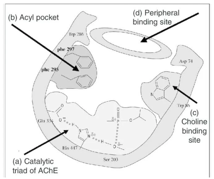
Diagram illustrating the active site of acetylcholinesterase (AChE), highlighting the catalytic triad, acyl pocket, and choline binding site.
Phosphodiesterase (PDE) Inhibitors
PDE inhibitors prevent the breakdown of second messengers, prolonging their physiological effects.
PDE3 Inhibitors (e.g., Milrinone): Known as "inodilators," they increase cAMP to improve cardiac contractility while simultaneously causing vasodilation to reduce preload and afterload.
PDE5 Inhibitors (e.g., Sildenafil): These selectively inhibit the breakdown of cGMP, making them effective pulmonary vasodilators and treatments for erectile dysfunction.
Nitric Oxide and Nitrovasodilators
Nitric Oxide (NO) is a potent endogenous vasodilator synthesized by endothelial cells.
Sodium Nitroprusside (SNP)
SNP is a rapid-acting donor that relaxes both arterial and venous smooth muscle.
Critical Warning: SNP carries a high risk of cyanide toxicity, especially at doses >2 mcg/kg/min. Signs include metabolic acidosis, altered mental status, and increased mixed venous oxygen content.
Nitroglycerin (NTG)
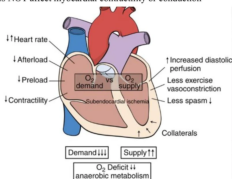
Unlike SNP, Nitroglycerin is an organic nitrate that primarily acts as a venodilator. By increasing venous capacitance, it reduces preload, which lessens myocardial wall tension and oxygen demand, making it ideal for treating angina.
Hydralazine
Hydralazine is a direct arterial vasodilator used for hypertension and pre-eclampsia. It is an effective afterload reducer but can cause reflex tachycardia due to sympathetic stimulation.
Other Specialized Agents
Endothelin Receptor Antagonists (e.g., Bosentan): These block the effects of endothelin, one of the body's most potent vasoconstrictors.
Catecholamine-Depleting Agents (e.g., Reserpine): These inhibit the uptake of catecholamines into storage vesicles, leaving them exposed to metabolic degradation.
Fenoldopam: A D1 agonist that provides renal vasodilation and natriuresis, making it useful in patients with renal insufficiency.
Diagram illustrating the relationship between myocardial oxygen demand and supply, and its impact on subendocardial ischemia.
Keep reading for free.
Create an account to finish this reading and make it part of your study routine.