An in-depth explanation of the cardiovascular system's structure and function, covering blood vessel types, their layers, endothelial and smooth muscle regulation, and the mechanisms of blood pressure control.
Understanding how the cardiovascular system maintains stability requires a deep dive into the structural components of blood vessels and the complex physiological mechanisms that regulate pressure. From the microscopic exchange of gases in capillaries to the systemic hormonal cascades that manage blood volume, every component plays a specific role in ensuring tissues receive adequate perfusion while maintaining hemodynamic stability.
The Architecture of the Vasculature
The circulatory system is composed of specialized vessels, each categorized by its primary physiological function: arterioles, capillaries, and veins.
Arterioles (Resistance Vessels): These vessels have a small radius and highly adjustable smooth muscle. They are the primary site of resistance, controlling the pressure drop and regulating the flow of blood into specific tissues.
Capillaries (Exchange Vessels): Characterized by a one-cell-thick wall and the greatest total surface area, capillaries are the primary site of nutrient and gas exchange. Their slow flow rate supports efficient diffusion.
Veins (Capacitance Vessels): With large, compliant lumens, veins act as blood reservoirs, containing the majority of the body's blood volume.
Blood Vessel Wall Layers
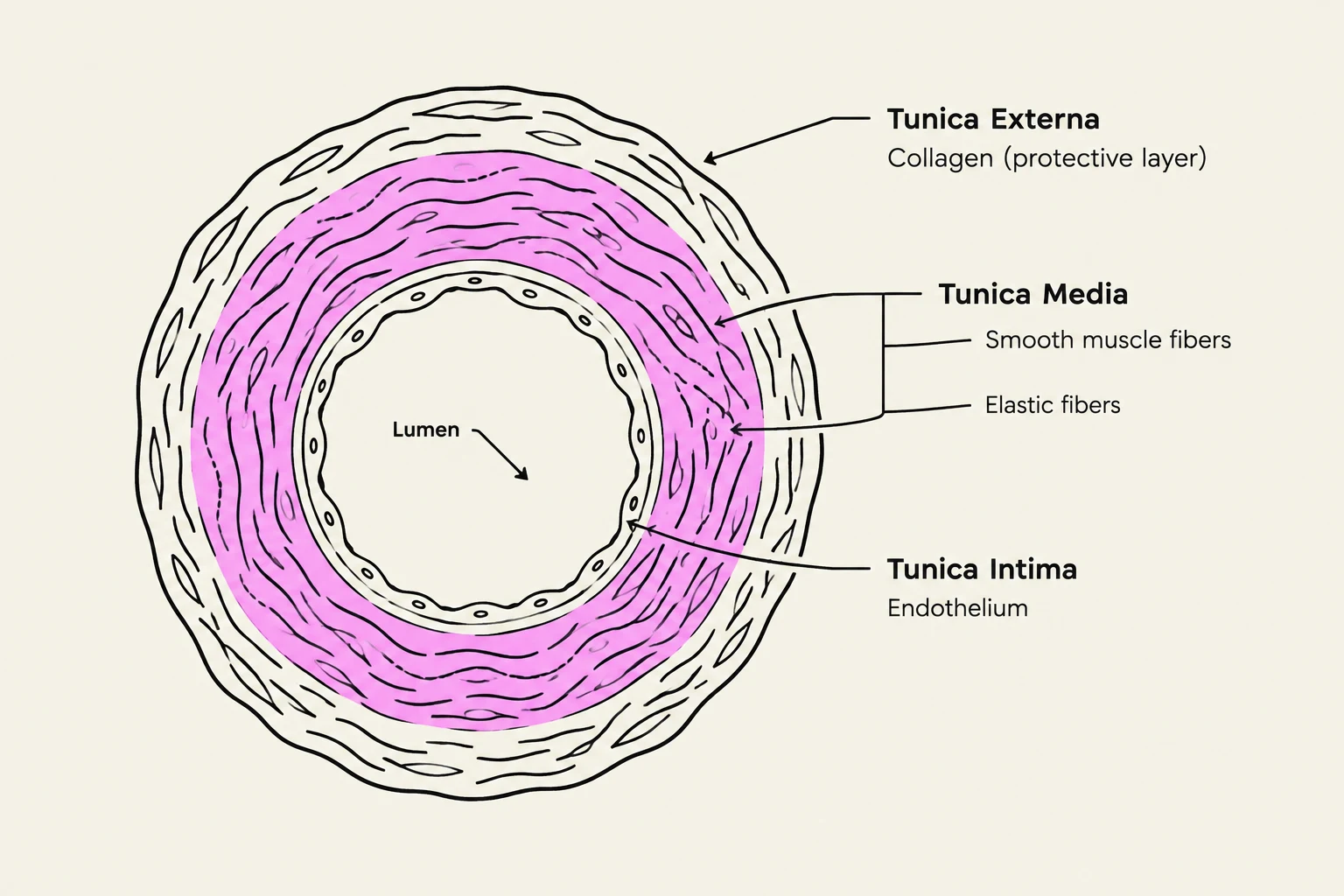
Diagram illustrating the cross-section of an artery, showing its three layers: Tunica Intima (endothelium), Tunica Media (smooth muscle fibers and elastic fibers), and Tunica Externa (collagen protective layer), surrounding the Lumen.
All blood vessels (excluding capillaries) are organized into three distinct layers, known as tunics:
Tunica Intima: The innermost layer, consisting of the endothelium and a basement membrane. It provides a smooth, selectively permeable surface that regulates vascular function.
Tunica Media: The middle layer, composed of circular smooth muscle and elastic fibers. This layer is responsible for changing vessel diameter through contraction and relaxation.
Tunica Externa: The outermost layer, made of collagen and elastin. It protects, strengthens, and anchors the vessel to surrounding tissues.
Endothelial and Smooth Muscle Regulation
The vascular endothelium is not merely a lining; it is a dynamic organ that maintains homeostasis by regulating vascular tone, platelet clotting, inflammation, hormone metabolism, and smooth-muscle growth.
Endothelial Homeostasis
Vascular tone is a delicate balance between vasodilators and vasoconstrictors. For example, Nitric Oxide (NO) promotes vasodilation, while Endothelin causes vasoconstriction. Endothelial dysfunction occurs when this balance shifts toward a pro-constrictive and pro-inflammatory state.
One critical pathway for relaxation is the eNOS–NO–sGC–cGMP–PKG pathway:
L-arginine is converted to NO by endothelial nitric oxide synthase (eNOS).
NO diffuses into vascular smooth muscle and activates soluble guanylyl cyclase (sGC).
sGC converts GTP to cGMP.
cGMP activates Protein Kinase G (PKG), which decreases intracellular Ca2+ and increases Myosin Light Chain Phosphatase (MLCP) activity.
This results in myosin-light-chain dephosphorylation and subsequent relaxation.
Smooth Muscle Contraction Mechanics
The diameter of a vessel is determined by the contraction or relaxation of the smooth muscle in the tunica media.
Contraction (Vasoconstriction): An increase in intracellular Ca2+ causes Ca2+ to bind to calmodulin. This complex activates Myosin Light Chain Kinase (MLCK), which phosphorylates the myosin light chain, triggering actin-myosin cycling and contraction. This decreases the lumen radius, increases resistance, and decreases local blood flow.
Relaxation (Vasodilation): A decrease in Ca2+ leads to decreased MLCK activity and increased MLCP activity, resulting in dephosphorylation and relaxation. This increases the lumen radius, decreases resistance, and increases local blood flow.
Exam Tip: Receptor Signaling
Pay close attention to the G-protein coupling of cardiovascular receptors:
α1: Gq protein →IP3/DAG→ Vasoconstriction.
α2: Gi protein →↓ cAMP.
β1: Gs protein →↑ cAMP →↑ Heart Rate/Contractility.
β2: Gs protein →↑ cAMP → Vasodilation.
M2: Gi protein →↓ Heart Rate.
M3: Gq protein → Vasoconstriction.
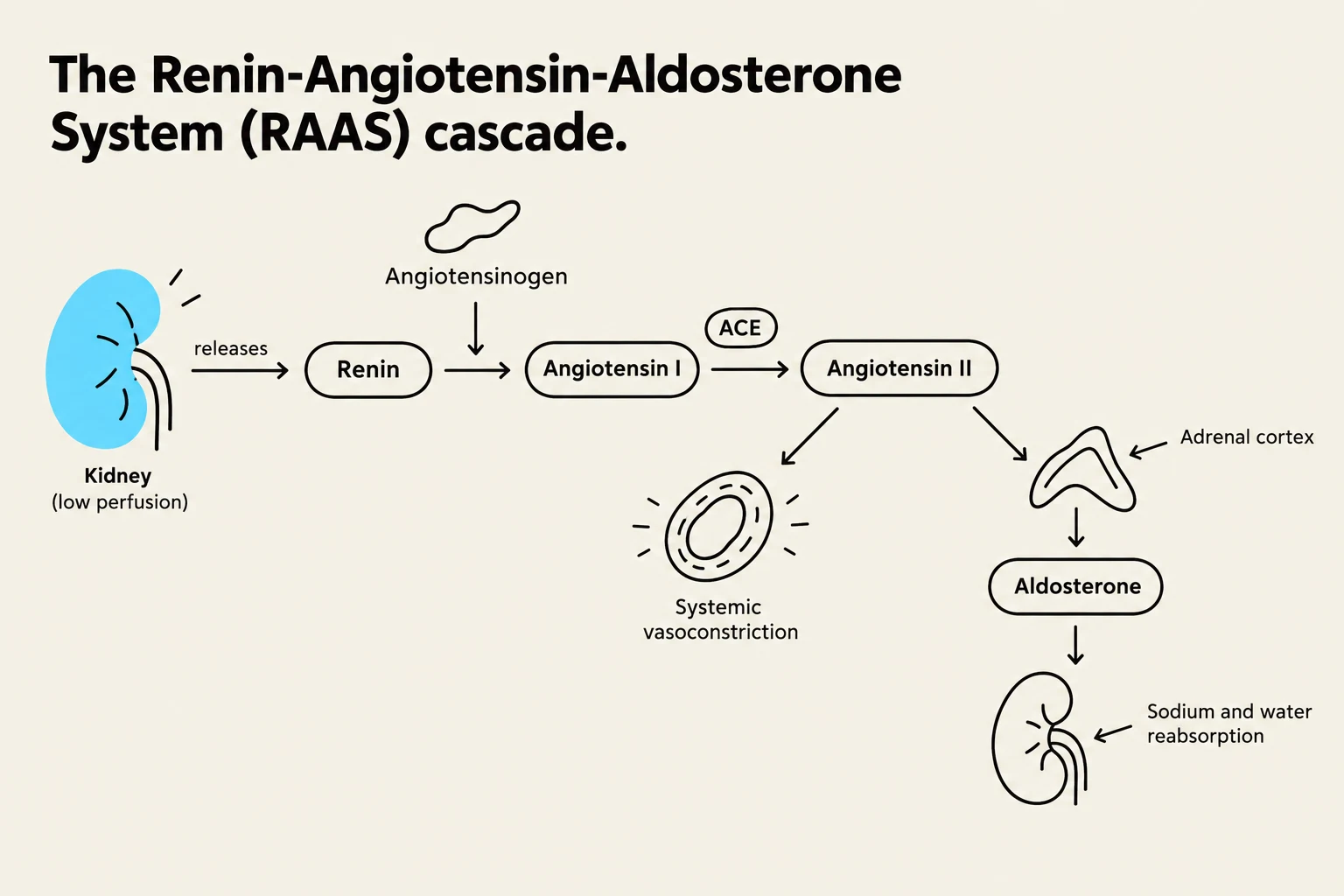
Diagram illustrating the Renin-Angiotensin-Aldosterone System (RAAS) cascade, showing the roles of the kidney, renin, angiotensinogen, angiotensin I, angiotensin II, ACE, adrenal cortex, and aldosterone in regulating blood pressure and fluid balance.
Hemodynamics and Blood Pressure Regulation
Blood pressure (BP) is the force exerted by blood against vessel walls. It is defined by three key metrics:
Systolic BP: The highest pressure during ventricular contraction.
Diastolic BP: The lowest pressure during ventricular relaxation.
Mean Arterial Pressure (MAP): The average pressure throughout the cardiac cycle.
The relationship between these variables is expressed by the following equations:
MAP≈DBP+31(SBP−DBP)MAP≈CO×SVR
Where CO is Cardiac Output and SVR is Systemic Vascular Resistance.
Mechanisms of Control
The body employs three distinct levels of regulation to maintain MAP:
Control Type
Timing
Major Mechanisms
Rapid
Seconds
Cardiovascular center, Autonomic Nervous System (ANS), Chemoreceptors
Baroreceptors sense changes in stretch. If BP increases, baroreceptor firing increases, leading to increased parasympathetic activity and decreased sympathetic activity, which lowers HR and contractility to reduce BP. Conversely, a drop in BP (such as when standing up) decreases baroreceptor firing, triggering a sympathetic surge to increase HR, contractility, and vasoconstriction.
Clinical Note: Orthostatic Hypotension
When a person stands suddenly, blood can pool in the lower body, reducing venous return, stroke volume (SV), and cardiac output (CO). If the baroreceptor reflex is inadequate, this results in orthostatic hypotension.
2. Long-term Regulation: The RAAS Pathway
The Renin-Angiotensin-Aldosterone System (RAAS) is a critical hormonal cascade for managing blood volume and long-term pressure.
Renin Release: Triggered by decreased renal perfusion, decreased NaCl delivery to the macula densa, or increased sympathetic stimulation of β1 receptors on juxtaglomerular (JG) cells.
Cascade: Renin converts angiotensinogen to Angiotensin I (Ang I), which is then converted to Angiotensin II (Ang II) by ACE.
Ang II Actions: Ang II is a potent vasoconstrictor (via AT1 receptors) and stimulates the release of Aldosterone.
Aldosterone Actions: Promotes Na+ reabsorption and $K^+$ excretion in the distal nephron, leading to water retention and increased blood volume.
Other hormonal players include ADH (increases water reabsorption) and ANP (promotes natriuresis and vasodilation to lower BP).
Pathophysiology of Hypertension
Hypertension can arise from various hemodynamic imbalances:
High Cardiac Output: Often driven by increased blood volume or sympathetic activity.
High SVR: Caused by persistent arteriolar vasoconstriction or vascular remodeling (wall thickening and lumen narrowing).
Renal/Hormonal: Driven by Na+ retention or excessive Ang II/Aldosterone activity.
Keep reading for free.
Create an account to finish this reading and make it part of your study routine.