The urinary system is a vital regulatory network responsible for filtering the blood, eliminating metabolic wastes—such as protein wastes, excess salts, and toxins—and maintaining the body's internal stability, known as homeostasis. Beyond simple waste removal, this system plays a critical role in managing fluid and blood volume, regulating pH and electrolyte balance, controlling blood pressure, and activating Vitamin D.
Organs of the Urinary System
The urinary system is composed of several key organs that work in sequence to filter, transport, store, and eliminate waste:
Kidneys (2): The primary filtration organs. They are retroperitoneal (located behind the peritoneum). Due to the presence of the liver, the right kidney sits slightly lower than the left.
Ureters (2): Smooth muscle tubes that use peristaltic waves to move urine from the kidneys to the bladder.
Urinary Bladder: A sterile, hollow, muscular sac that serves as a reservoir for urine. It can hold approximately 300–500 mL of urine.
Urethra: The tube through which urine passes to the outside of the body. In women, it is approximately 1.5 inches long, whereas in men, it is about 8 inches long and passes through the prostate and penis.
Internal Kidney Anatomy
The kidney is highly vascular and organized into distinct regions:
Renal Cortex: The outer portion of the kidney.
Renal Medulla: The middle portion, which contains structures like the renal pyramids.
Renal Pelvis: A funnel-like structure that collects urine before it enters the ureter.
Hilum: The notch on the kidney where blood vessels, nerves, and the ureter enter and exit.
Renal Blood Flow: The Filtration Pathway
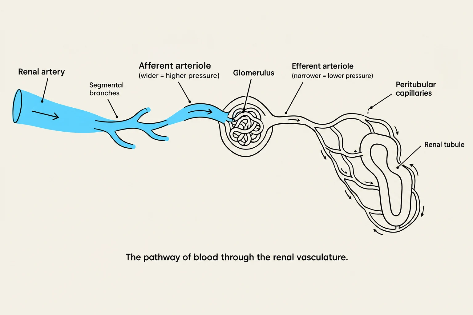
Diagram illustrating the pathway of blood through the renal vasculature, including the renal artery, afferent arteriole, glomerulus, efferent arteriole, peritubular capillaries, and renal tubule.
The kidneys are incredibly efficient, receiving approximately 20–25% of the total cardiac output. This high volume of blood is necessary to constantly filter the body's plasma.
The blood follows a precise pathway through the renal vasculature:
Afferent Arteriole: This vessel delivers blood to the glomerulus. It has a wider diameter to maintain the high pressure required for filtration.
Glomerulus: A knotted cluster of capillaries where the actual filtration occurs.
Efferent Arteriole: Blood exits the glomerulus through this vessel, which has a narrower diameter than the afferent arteriole.
Peritubular Capillaries: These surround the renal tubules and facilitate the exchange of substances through reabsorption and secretion.
Venous Return: Filtered blood travels from the peritubular capillaries → venules → cortical radiate veins → arcuate veins → interlobar veins →renal vein, which finally empties into the inferior vena cava.
The Nephron: The Functional Unit of the Kidney
Each kidney contains approximately one million nephrons, which are the microscopic structures responsible for forming urine. There are two primary types of nephrons:
Cortical Nephrons: These have glomeruli located in the outer cortex and possess short loops of Henle.
Juxtamedullary Nephrons: These have glomeruli located at the corticomedullary border.
The Process of Urine Formation
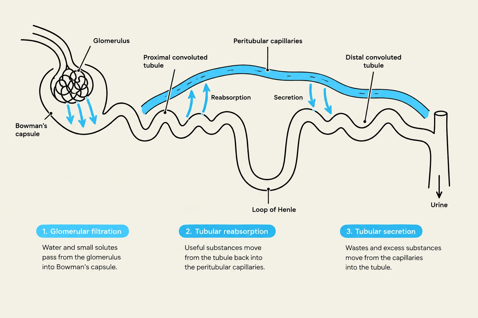
Diagram illustrating the processes of glomerular filtration, tubular reabsorption, and tubular secretion in the nephron of the kidney.
Urine formation occurs through three distinct physiological processes:
Glomerular Filtration: This occurs within the renal corpuscle, which consists of the glomerulus and the surrounding Bowman’s capsule. High pressure forces water and small solutes (ions, glucose, urea, creatinine) across the filtration membrane into the capsule. Crucially, large substances like Red Blood Cells (RBCs), White Blood Cells (WBCs), and large proteins (like albumin) do not pass through. The resulting fluid is called glomerular filtrate.
Tubular Reabsorption: As the filtrate moves through the proximal convoluted tubule (PCT), loop of Henle, and distal convoluted tubule (DCT), the body reclaims necessary substances (water, glucose, amino acids, and electrolytes) by moving them from the tubules back into the peritubular capillaries.
Tubular Secretion: This is the reverse of reabsorption; substances are moved from the capillaries and interstitial fluid into the tubules to be eliminated.
Clinical Note: GFR
The Glomerular Filtration Rate (GFR) is a key indicator of renal function. It measures how fast renal clearance is occurring. A normal GFR for an adult is approximately 125 mL/min.
Homeostasis and Endocrine Functions
The kidneys are not just filters; they are endocrine organs that secrete hormones to maintain systemic balance.
Blood Pressure and the RAAS Pathway
When the kidneys detect low blood pressure, low blood volume, or low sodium levels, they initiate the Renin–Angiotensin–Aldosterone System (RAAS):
Renin: An enzyme released by the kidneys that converts angiotensinogen (from the liver) into Angiotensin I.
Angiotensin II: Angiotensin I is converted to Angiotensin II by Angiotensin-Converting Enzyme (ACE), primarily in the lungs. Angiotensin II is a strong vasoconstrictor that raises blood pressure and stimulates the adrenal glands.
Aldosterone: Released by the adrenal glands, this hormone increases sodium reabsorption in the distal convoluted tubule and collecting duct. Because water follows sodium via osmosis, this increases blood volume and blood pressure.
Other hormonal regulators include:
Anti-diuretic Hormone (ADH): Secreted by the posterior pituitary, it increases water reabsorption in the tubules, which decreases urine volume and increases urine concentration.
Atrial Natriuretic Peptide (ANP): Secreted by the heart atria when blood volume is too high, it suppresses ADH and aldosterone to increase the excretion of water and sodium, thereby decreasing blood volume.
Other Homeostatic Roles
Erythropoietin (EPO): When the kidneys detect hypoxia (low oxygen), they secrete EPO to stimulate the formation of red blood cells (erythropoiesis).
Vitamin D Activation: The kidneys convert Vitamin D into its metabolically active form (calcitriol), which is essential for calcium balance.
Acid-Base and Electrolyte Balance: Through the regulation of ion secretion and reabsorption in the distal tubules, the kidneys maintain the body's pH and electrolyte levels.
Storage and Elimination
Once urine is formed in the collecting ducts, it flows into the renal pelvis and down the ureters to the urinary bladder.
Micturition vs. Incontinence
The process of urinating is known as micturition (or voiding). During this process, the detrusor muscle of the bladder contracts, and the involuntary internal sphincter relaxes.
Normal Micturition: A coordinated process of bladder contraction and sphincter relaxation.
Incontinence: The involuntary loss of urine, which can occur due to various medical conditions.
Aging and the Urinary System
As humans age, several physiological changes occur in the urinary system:
A decrease in the total number of functioning nephrons.
Loss of bladder muscle tone, which can lead to increased frequency or inefficient emptying.
Increased risk of Urinary Tract Infections (UTIs) due to residual urine.
Increased nocturia (waking up at night to urinate) due to decreased bladder capacity.
Keep reading for free.
Create an account to finish this reading and make it part of your study routine.