An overview of the human urinary system, detailing its components, kidney anatomy, nephron structure and function, and essential kidney functions in maintaining homeostasis.
The human body is a complex environment that requires constant monitoring to stay healthy. To maintain homeostasis—the stable internal state necessary for survival—the body must precisely regulate its chemical composition, fluid levels, and blood pressure. The urinary system serves as the body's primary filtration and regulatory plant, ensuring that metabolic wastes are removed while vital nutrients, ions, and water are conserved.
The Urinary System Components
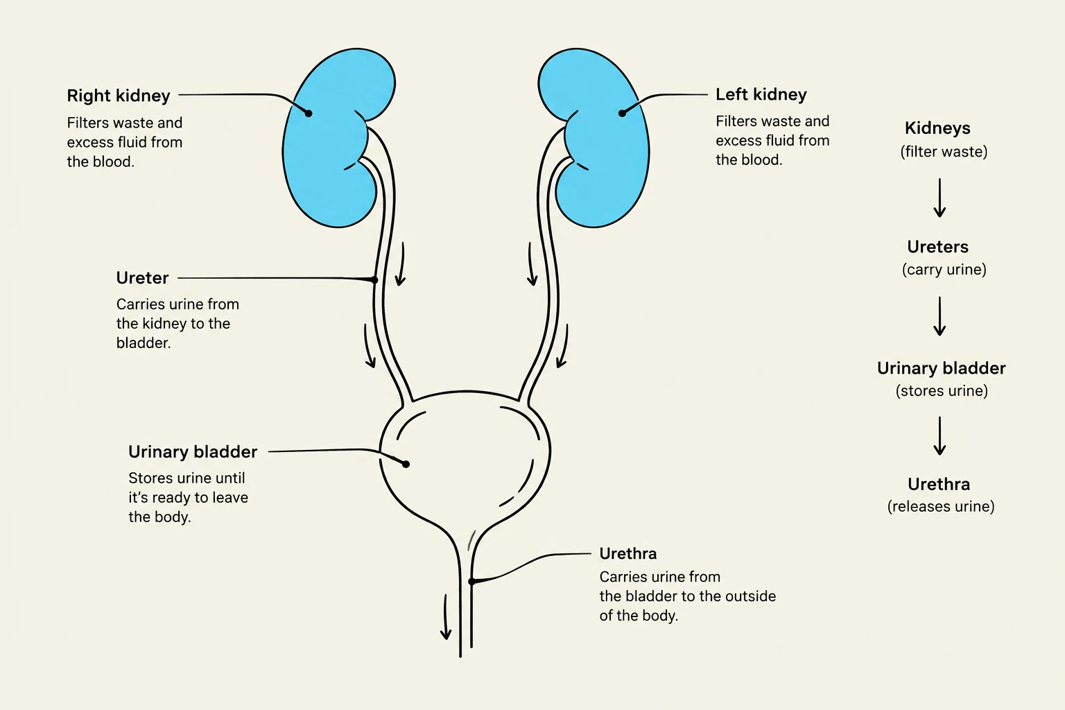
The urinary system is a coordinated network of organs designed to filter blood, produce urine, and eliminate waste from the body. It consists of four primary components:
Kidneys: The functional powerhouses that filter the blood and form urine.
Ureters: Tubes that transport urine from the kidneys to the bladder.
Urinary Bladder: A muscular sac that can stretch to hold up to two cups of urine.
Urethra: The channel through which urine is finally eliminated from the body.
Diagram of the human urinary system, illustrating the kidneys, ureters, urinary bladder, and urethra, along with their functions.
Kidney Anatomy: Structure and Protection
The kidneys are paired, bean-shaped organs located along the posterior abdominal wall, a position known as being retroperitoneal. Interestingly, the right kidney typically sits slightly lower than the left. On the medial side of each kidney is an indentation called the renal hilum, which serves as the entry and exit point for blood vessels, nerves, and the ureter.
To ensure stability and protection, the kidneys are encased in several layers:
Fibrous capsule: A thin, dense irregular connective tissue layer that maintains the kidney's shape.
Perinephric fat (adipose capsule): Provides cushioning and support.
Renal fascia: A layer of dense irregular connective tissue that anchors the kidney to surrounding structures.
Clinical Note: Renal Ptosis
If a person experiences a significant loss of supporting adipose tissue, the kidney may undergo "renal ptosis" (nephroptosis), an abnormal downward displacement. This can cause the ureter to kink, leading to urine backup and a condition called hydronephrosis, where the renal pelvis dilates.
Internal Organization: Cortex and Medulla
When looking at a cross-section of the kidney, the functional tissue (parenchyma) is divided into two main regions:
Renal Cortex: The outer region of the kidney.
Renal Medulla: The inner region, which contains renal pyramids.
The renal pyramids are subdivided by extensions of the cortex known as renal columns. At the tip of each pyramid is the renal papilla, which drains urine into the minor calyx.
The Nephron: The Functional Unit of Filtration
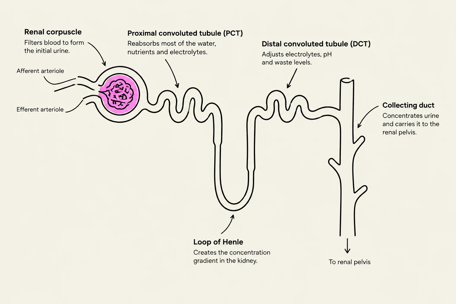
The actual work of the kidney is performed by microscopic units called nephrons. Each nephron consists of two main parts: the renal corpuscle and the renal tubule.
The Renal Corpuscle
Filtration begins in the renal corpuscle, which contains the glomerulus (a capillary network) surrounded by the glomerular (Bowman's) capsule. Blood enters via the afferent arteriole and exits through the efferent arteriole. The fluid forced out of the blood into the capsular space is called filtrate.
The Renal Tubule and Urine Formation
The filtrate travels through several specialized segments of the renal tubule, each with a specific role in modifying the fluid:
Proximal Convoluted Tubule (PCT): The site of "bulk reabsorption". It uses microvilli to increase surface area and reabsorbs approximately 65% of filtered sodium and water, as well as nearly all glucose and amino acids.
Nephron Loop (Loop of Henle): This segment establishes an osmotic gradient. The descending limb is permeable to water but not solutes, making the filtrate more concentrated. The ascending limb is impermeable to water but permeable to solutes (like NaCl), making the filtrate more dilute.
Distal Convoluted Tubule (DCT): This area performs "fine-tuning" of the filtrate, reabsorbing ions like Na+ and $Cl^-$.
Collecting Duct: While not technically part of a single nephron, it receives fluid from many nephrons to perform the final adjustment of urine composition.
Diagram illustrating the structure and function of a nephron, the functional unit of the kidney, including the renal corpuscle, proximal convoluted tubule, loop of Henle, distal convoluted tubule, and collecting duct.
Essential Kidney Functions
The primary goal of the kidney is not merely "making urine," but rather regulating the composition of the blood to maintain homeostasis.
1. Elimination of Metabolic Wastes
The kidneys prevent toxic levels of metabolic byproducts from building up in the blood. Key wastes include urea, uric acid, and creatinine.
2. Regulation of Ions and Acid-Base Balance
The kidneys maintain the delicate balance of electrolytes, such as sodium (Na+), potassium ($K^+$), and calcium (Ca2+). They also regulate blood pH by altering the levels of hydrogen ($H^+$) and bicarbonate (HCO3−) ions.
3. Water Balance and Blood Pressure
By controlling how much water is retained versus lost in urine, the kidneys directly influence blood volume. Because blood volume dictates blood pressure, this is a critical regulatory mechanism.
4. Endocrine Functions
The kidneys act as endocrine glands by producing and releasing several vital substances:
Renin: Released by the juxtaglomerular (JG) apparatus when blood pressure drops. Renin activates a pathway to increase blood pressure.
Erythropoietin (EPO): Released when blood oxygen levels are low to stimulate the production of red blood cells (erythrocytes).
Calcitriol: The active form of Vitamin D, which increases calcium absorption from the intestines to raise blood calcium levels.
The Juxtaglomerular Apparatus (JGA): The Sensor
The kidney has an internal "monitoring system" called the Juxtaglomerular Apparatus (JGA), located where the DCT returns to the vascular pole of its own renal corpuscle. It consists of two main components:
Granular (JG) cells: Modified smooth muscle cells in the afferent arteriole that sense changes in blood pressure and release renin.
Macula densa: Specialized cells in the DCT that sense the concentration of NaCl in the tubular fluid.
How the JGA Works: A Logic Check
Low NaCl delivery to Macula Densa: This often signals a low Glomerular Filtration Rate (GFR) or low blood flow. The Macula densa signals the JG cells to increase renin release to restore pressure.
High NaCl delivery to Macula Densa: This signals a high GFR or high flow. The response is to decrease renin release to help lower the GFR.
Diagram illustrating the juxtaglomerular apparatus, including the glomerulus, Bowman's capsule, distal convoluted tubule (DCT), afferent and efferent arterioles, granular cells, and macula densa cells. It also depicts the signaling pathway involving the renin-angiotensin system.
Keep reading for free.
Create an account to finish this reading and make it part of your study routine.