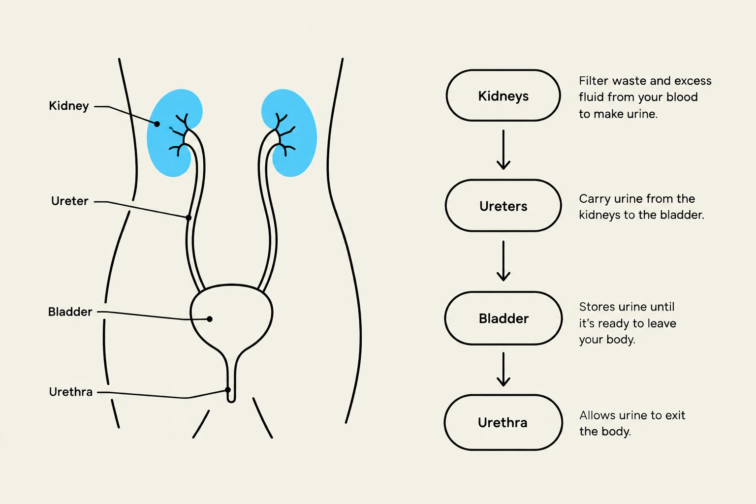
Effective urinary elimination management is a cornerstone of nursing care, requiring a deep understanding of renal anatomy, diagnostic procedures, and sterile techniques. The urinary system—comprising the kidneys, ureters, bladder, and urethra—works to filter blood, regulate electrolytes, and excrete waste. For the nurse, managing this system involves more than just monitoring output; it requires precise specimen collection, the prevention of infections, and the careful management of various urinary diversions and catheters.
Specimen Collection and Diagnostics
Accurate diagnosis begins with high-quality specimens. The method of collection depends entirely on the clinical question being asked.
Methods of Collection
Routine Voided Sample: Collected in a clean container for urinalysis to assess concentration and abnormal constituents. Patients should be instructed to avoid contamination from stool or tissue.
Clean-Catch Midstream: This is the gold standard for culture and sensitivity (C&S) to identify specific organisms and determine antibiotic susceptibility. To perform this, the patient cleanses the meatus, begins voiding into the toilet, and then catches the "midstream" portion in a sterile container.
Catheter Specimens: For patients with indwelling catheters, urine must be collected from the designated sampling port using aseptic technique. Never collect a culture from the drainage bag, as the urine there is not fresh and may contain colonizing bacteria rather than the actual infecting organism.
Straight Catheter Specimen: In acute care settings, a sterile straight catheter may be used to obtain a fresh sample.
Timed Collection: This requires collecting every drop of urine produced during a specific window. At the start time, the patient voids and that urine is discarded; all subsequent voids are collected, including the final void at the end of the timed period.
Exam Tip: If a timed specimen is accidentally missed or discarded, do not attempt to estimate the volume or add water. Contact the laboratory to follow specific restart instructions.
Diagnostic Tools
Beyond urine samples, nurses utilize several tools to assess urinary health:
Bladder Scans: A noninvasive ultrasound used to estimate bladder volume and postvoid residual.
Cystoscopy: Provides direct visualization of the urethra and bladder.
Ultrasound and CT Scans: Used to evaluate urinary structures, stones, or obstructions.
Diagram illustrating the human urinary system, including the kidneys, ureters, bladder, and urethra, with descriptions of their functions.
Catheterization and Drainage Systems
Catheters are indicated for clients who are unable to void or require constant monitoring of fluid status. Because of the high risk of infection, most catheters should be temporary.
Catheter Types
Catheter size is measured in French (Fr), which describes the external diameter.
Straight (Intermittent) Catheter: Used for one-time drainage; it has no balloon.
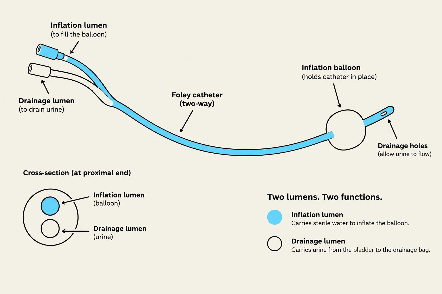
Two-way Foley: Contains one lumen for drainage and one for the balloon.
Three-way Foley: Includes an additional lumen for bladder irrigation.
Coudé Catheter: Features a curved tip to assist with difficult catheterizations, such as in patients with enlarged prostates.
External Devices: Condom catheters (for males) and external female devices can collect urine without entering the bladder. However, it is critical to remember that external devices do not relieve urinary retention.
Sterile Insertion Technique
Insertion requires a strict sterile field and a provider's order.
Preparation: Verify the order, identity, and allergies. Establish a sterile field and apply sterile gloves.
Insertion: For females, use the non-dominant hand to expose the meatus and cleanse with an approved solution. For males, hold the penis and cleanse with swabs.
Advancement: Once a "flash" of urine is seen in the tubing, advance the catheter an additional 2–3 inches to ensure the tip is inside the bladder before inflating the balloon.
Inflation: Inflate the balloon only with the specific volume and fluid type labeled on the catheter.
Diagram illustrating the components and functions of a two-way Foley catheter, including its inflation lumen, drainage lumen, and balloon.
Preventing and Managing Infection (CAUTI)
Catheter-Associated Urinary Tract Infections (CAUTIs) are a significant risk. Prevention relies on maintaining a closed, unobstructed drainage system.
Nursing Interventions for Prevention
Hygiene: Provide routine perineal hygiene (soap and water) at least once per shift.
Positioning: Always keep the drainage bag below the level of the bladder to prevent reflux and ensure it remains off the floor.
Maintenance: Avoid kinks in the tubing and ensure the system remains closed.
Removal: Review the necessity of the catheter daily and remove it promptly once it is no longer clinically indicated.
Urinary Diversions and Specialized Access
When the normal urinary tract is bypassed or surgically altered, specialized devices are used:
Suprapubic Catheter: Enters the bladder through the abdomen, bypassing the urethra.
Ileal Conduit (Urostomy): Urine drains through a segment of the intestine to a stoma. The nurse must assess the stoma; it should appear pink, moist, and meaty. Discoloration or a lack of output must be reported immediately.
Nephrostomy: Drains urine directly from the kidney. Monitor the insertion site, output, and avoid any traction (pulling) on the tubing.
Pharmacology and Clinical Indicators
Monitoring Output and Labs
A critical indicator of renal perfusion is a minimum urine output of 30 mL/hour. Nurses must monitor the following laboratory values to assess kidney function and electrolyte balance:
Lab Test
Normal Reference Range
Clinical Significance
BUN
7–20 mg/dL
Kidney function/waste product
Creatinine
0.6–1.3 mg/dL
Primary indicator of kidney function
Sodium (Na+)
135–145 mEq/L
Fluid/electrolyte balance
Potassium (K+)
3.5–5.0 mEq/L
Electrolyte balance; critical for heart/muscle
Medications Affecting Elimination
Diuretics (e.g., Furosemide): Increase urine output. Monitor for dehydration, low blood pressure, and especially potassium loss.
Anticholinergics (e.g., Oxybutynin): Used to reduce bladder urgency.
Opioids and Antihistamines: These can cause urinary retention.
Clinical Reasoning: A Worked Example
Scenario: A postoperative patient has not voided for 6 hours. They report lower abdominal discomfort. A bladder scan shows a volume of 650 mL.
Analysis:
Identify the Problem: The patient is experiencing urinary retention.
Cues: The discomfort and the high bladder scan volume (650 mL) are the primary cues.
Priority Action: The nurse should follow the facility's retention protocol, which may include notifying the provider or preparing for intermittent catheterization to empty the bladder.
Nursing Diagnosis Tip: When writing an "Actual" diagnosis, use the format: Problem + Related Factor + Signs/Symptoms.
Example: Urinary retention related to reduced bladder contractility after anesthesia as evidenced by inability to void and a bladder scan of 650 mL.
Keep reading for free.
Create an account to finish this reading and make it part of your study routine.