Understanding Ventilation, Perfusion, and V/Q Mismatch in Respiratory Health
This content explains the fundamental concepts of ventilation and perfusion in the respiratory system, detailing V/Q mismatch and its causes. It also covers traumatic lung injuries like flail chest, and complications such as pneumothorax and pleural effusion, providing a comprehensive overview of respiratory mechanics
To understand how our body maintains life, we must look at the seamless partnership between the respiratory and cardiovascular systems. Oxygenation is not just about breathing; it is about the successful delivery of air to the lungs and the subsequent delivery of blood to those same areas to pick up that oxygen. When this partnership is disrupted, the resulting imbalance can lead to life-threatening conditions ranging from simple shortness of breath to severe tissue damage.
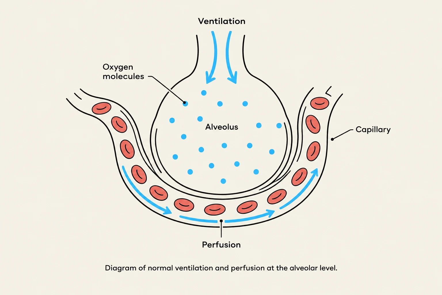
Diagram of normal ventilation and perfusion at the alveolar level.
Ventilation vs. Perfusion: The Fundamentals
The process of oxygenation relies on two distinct but interdependent mechanisms: Ventilation and Perfusion.
Ventilation (V): This refers to the mechanical process of air moving in and out of the lungs. For ventilation to be effective, the airways must remain clear and open so that fresh, oxygen-rich air can reach the alveoli (the tiny air sacs in the lungs).
Perfusion (Q): This refers to the movement of blood through the pulmonary capillaries that surround the alveoli. The purpose of perfusion is to allow the blood to "pick up" the oxygen from the alveoli and circulate it throughout the rest of the body.
In a healthy individual, these two processes work "hand in hand." Air enters the alveoli, and blood flows past them to facilitate gas exchange.
V/Q Mismatch: When the Partnership Fails
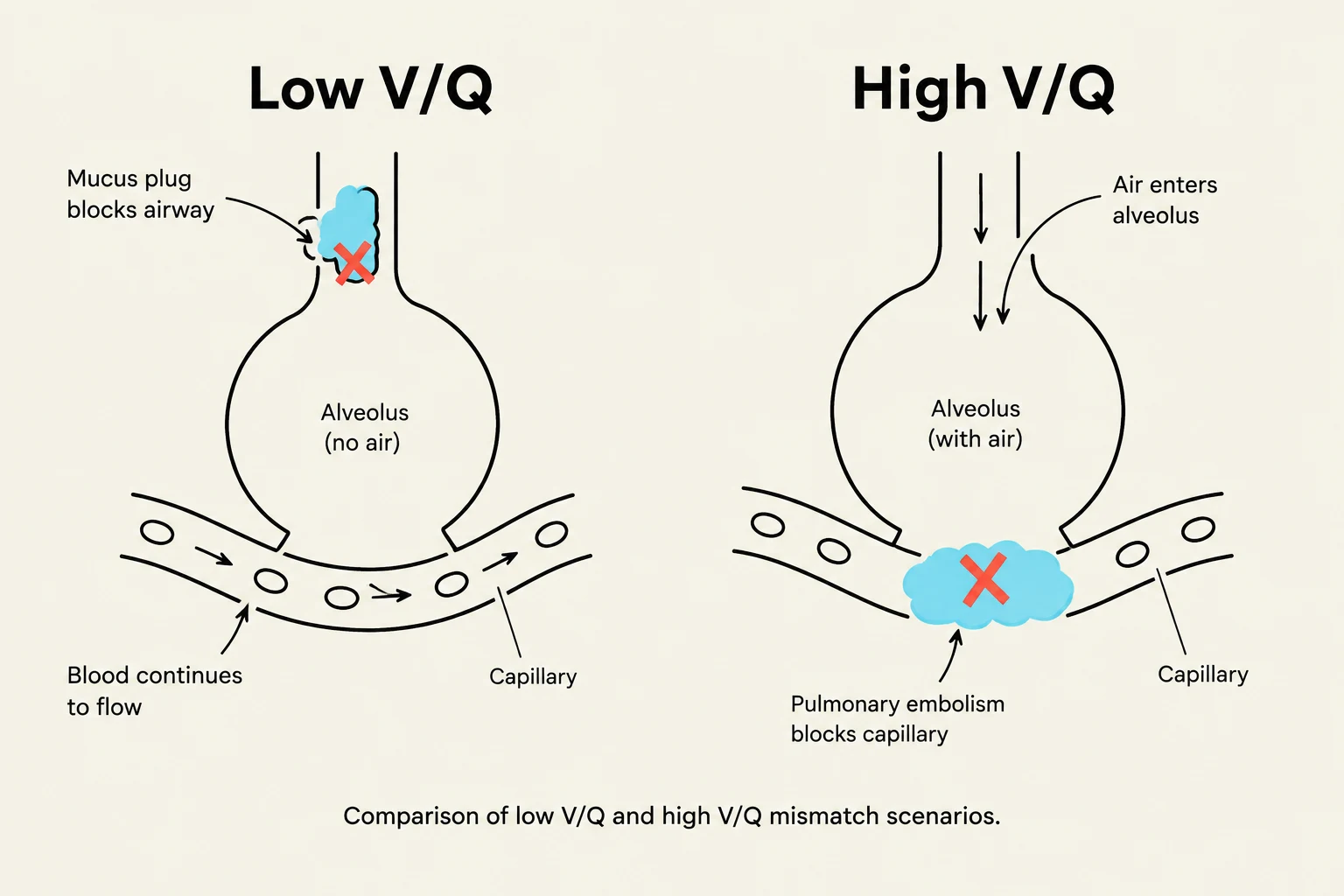
This diagram illustrates two scenarios of ventilation-perfusion (V/Q) mismatch in the lungs: low V/Q due to a mucus plug blocking an airway, and high V/Q due to a pulmonary embolism blocking a capillary.
A V/Q mismatch occurs when the amount of air reaching the alveoli (Ventilation) does not match the amount of blood reaching those same alveoli (Perfusion). This mismatch is the primary driver behind oxygenation failures.
1. Low V/Q Mismatch (Ventilation Problem)
In a low V/Q scenario, perfusion is present, but ventilation is impaired. The blood is coming to the alveoli, but there isn't enough oxygen available for it to pick up. Common causes include:
Pulmonary Edema: Fluid in the alveoli prevents oxygen from crossing into the blood.
Physical Obstruction: Choking or mucus production (as seen in asthma or bronchitis) blocks the airway.
Atelectasis: The collapse of the alveoli themselves.
2. High V/Q Mismatch (Perfusion Problem)
In a high V/Q scenario, ventilation is working perfectly—air is moving in and out of the lungs easily—but the blood flow is blocked or diminished.
Pulmonary Embolism: This is the most common cause of high V/Q mismatch (accounting for approximately 90% of cases). A clot blocks the blood vessel, so even though the alveoli are full of oxygen, there is no blood there to collect it.
Exam Tip: Hypoxemia vs. Hypoxia
It is crucial to distinguish between these two terms. Hypoxemia refers specifically to low oxygen levels in the blood. Hypoxia refers to decreased oxygen delivery to the tissues. Hypoxemia will almost always lead to hypoxia, which can ultimately result in ischemia (tissue damage due to lack of blood/oxygen).
Traumatic Lung Injury: Flail Chest
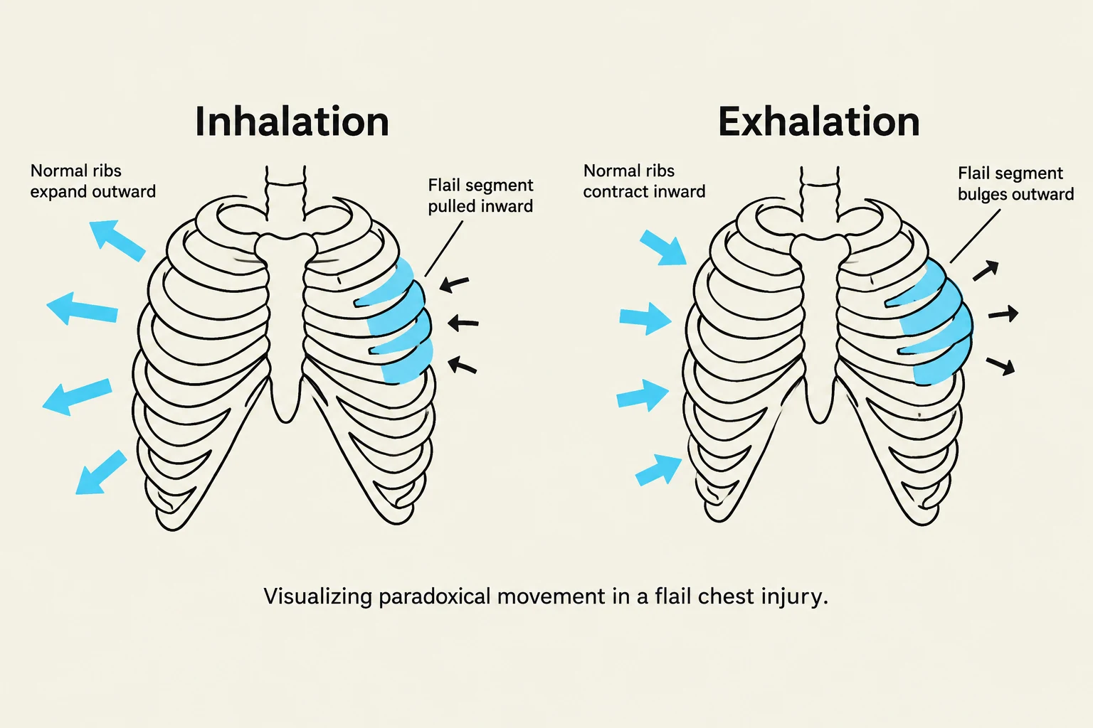
This diagram illustrates the paradoxical movement of a flail chest segment during inhalation and exhalation.
Trauma can cause severe mechanical disruptions to the respiratory system. One of the most significant injuries is a flail chest.
Definition and Mechanism
A flail chest is defined by two or more consecutive rib fractures in two or more places. This creates a "free-floating" segment of the rib cage that is no longer structurally connected to the rest of the chest wall.
Paradoxical Movement
The hallmark sign of a flail chest is paradoxical movement. In normal breathing, the chest expands when you inhale and contracts when you exhale. In a patient with a flail chest:
When inhaling: The free-floating segment is sucked inward toward the chest cavity.
When exhaling: The segment bulges outward.
Clinical Presentation and Treatment
Patients often experience intense pain, leading to very shallow, ineffective breathing. This can cause tachypnea (fast breathing) and tachycardia (fast heart rate) as the body struggles to compensate.
Treatment Approaches:
Supportive Care: Monitoring and providing oxygen therapy (such as CPAP or BiPAP) to provide positive pressure and push oxygen into the lower lung bases.
Surgical Intervention: In some cases, surgery is required to fix the broken bones.
Note on Historical Practice: We no longer "wrap" the chest to immobilize broken ribs, as this inhibits deep breathing and worsens the condition.
Complications: Pneumothorax and Pleural Effusion
Trauma or underlying disease can lead to air or fluid accumulating in the pleural space (the area between the lung and the chest wall).
Pneumothorax (Air in the Pleural Space)
A pneumothorax occurs when the negative pressure that keeps the lungs inflated is lost.
Open Pneumothorax: Acts like a "two-way valve" where air can enter and escape.
Tension Pneumothorax: Acts like a "one-way valve." Air enters the pleural space but cannot get out. This causes pressure to build, eventually collapsing the lung and pushing the heart and trachea to the opposite side (tracheal deviation). This is a medical emergency that can lead to cardiac arrest.
Pleural Effusion (Fluid in the Pleural Space)
While a pneumothorax involves air, a pleural effusion involves the accumulation of fluid. The type of fluid can indicate the cause:
Transudative: Clear, watery fluid (often seen in overhydration or heart failure).