Uterine sarcomas are rare and aggressive malignancies arising from the mesenchymal components of the uterus. Unlike carcinomas, which originate from the epithelial lining, sarcomas develop from the smooth muscle (leiomyosarcoma), connective tissue, or vascular structures. Their rarity, diverse histological subtypes, and aggressive nature present significant diagnostic and therapeutic challenges. This article provides an overview of the pathology, molecular analysis, imaging modalities, and therapeutic strategies for uterine sarcomas, aiming to equip clinicians with essential knowledge for managing these complex cases.
Pathology and Molecular Analysis
Accurate diagnosis of uterine sarcoma hinges on meticulous histopathological examination and, increasingly, on molecular profiling to identify specific drivers and prognostic markers.
Uterine Tumor Resembling Ovarian Sex Cord Tumor (UTROSCT)
UTROSCT is a rare subtype characterized by a proliferation of bland spindle cells with features suggestive of ovarian sex cord differentiation. Molecular analysis has identified recurrent gene fusions, particularly involving $ESR1$ or $GREB1$, which are crucial for its classification.
Rhabdomyosarcoma (RMS)
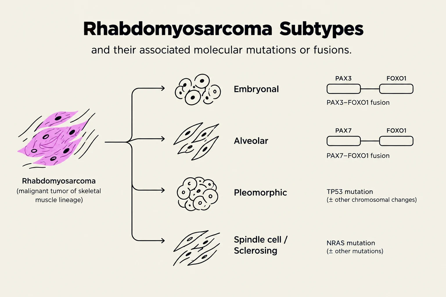
Rhabdomyosarcoma is a malignant mesenchymal tumor characterized by skeletal muscle differentiation. It is further classified into several subtypes, each with distinct molecular features:
Embryonal RMS: Often associated with $DICER1$ mutations.
Pleomorphic RMS: Frequently exhibits $PIK3CA$ and $TP53$ mutations.
Alveolar RMS: Linked to $FOXO1$ fusions.
Immunohistochemical staining for myogenin or MyoD1 is essential for confirming the diagnosis.
This diagram illustrates the subtypes of rhabdomyosarcoma, a malignant tumor of skeletal muscle lineage, and their associated molecular mutations or fusions. The subtypes shown are Embryonal, Alveolar, Pleomorphic, and Spindle cell/Sclerosing, each linked to specific genetic alterations like PAX3-FOXO1 fusion, PAX7-FOXO1 fusion, TP53 mutation, or NRAS mutation.
Müllerian Adenosarcoma (MAS)
Müllerian adenosarcoma is a biphasic tumor composed of a benign epithelial component and a malignant mesenchymal (stromal) component. High-grade features and a sarcomatous component exceeding 25% are associated with a poorer prognosis.
Other Sarcoma Entities and Molecular Findings
Other important uterine sarcoma subtypes include high-grade endometrial stromal sarcomas, which may harbor BCOR internal tandem duplications, and specific leiomyosarcomas that can show $PLAG1$ rearrangements or MDM2/CDK4 amplifications. Molecular testing is increasingly vital for identifying actionable targets and understanding tumor biology.
Imaging Protocols
Imaging plays a critical role in the initial workup, staging, and surveillance of uterine sarcomas.
Initial Workup
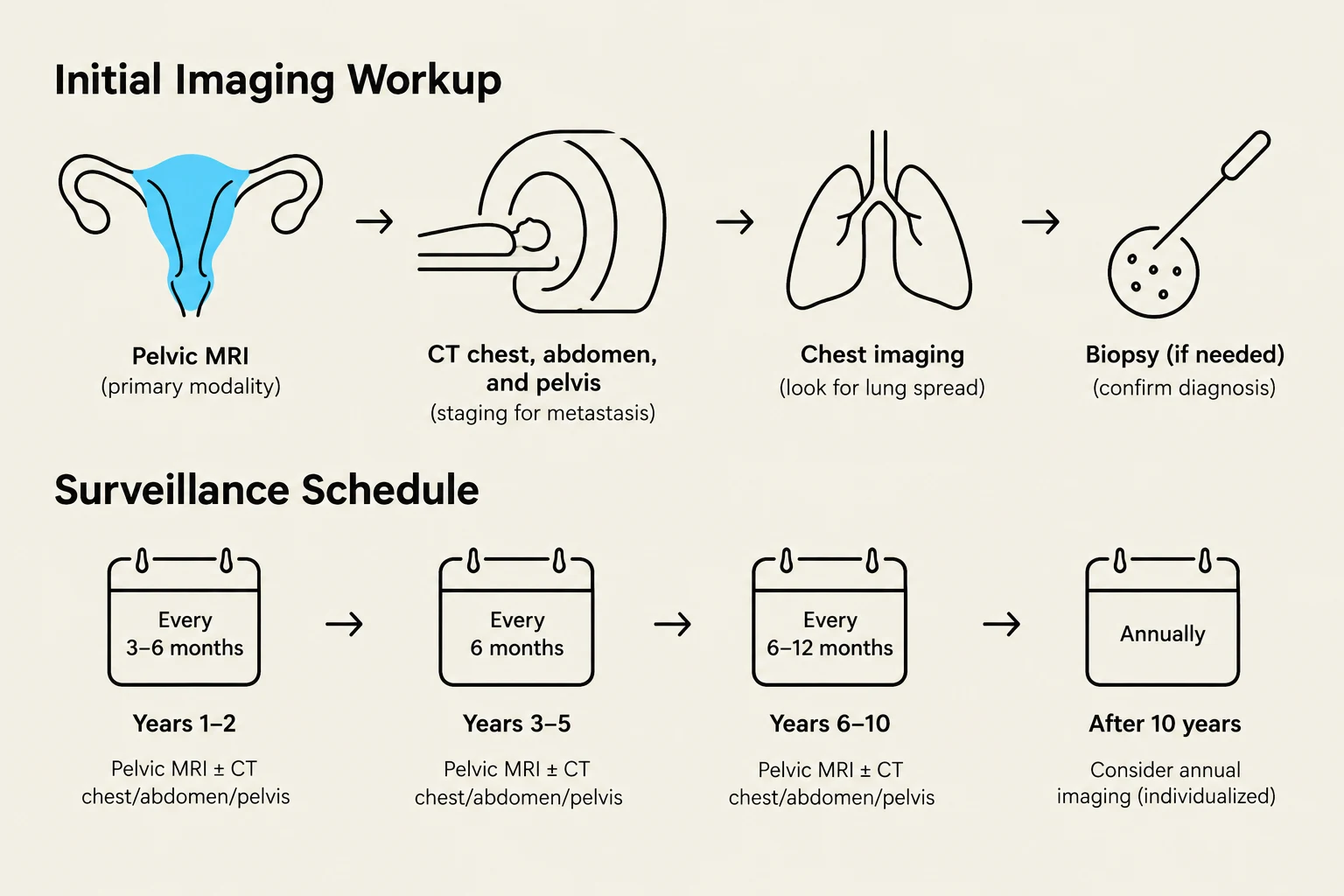
The initial imaging evaluation typically includes:
Chest/Abdomen/Pelvis CT: Performed with contrast, except for screening chest CT, to assess for metastatic disease.
Pelvis MRI: Utilized with and without contrast to evaluate local tumor extension, involvement of adjacent structures, and to assess for residual disease after surgery, particularly in cases of incomplete resection.
FDG-PET/CT: Reserved for clarifying ambiguous findings on conventional imaging or when there is a high suspicion of metastasis.
Surveillance
Following initial treatment, patients are typically monitored with serial imaging to detect recurrence or metastasis.
Chest/Abdomen/Pelvis CT: Generally performed every 3–6 months for the first 3 years, then transitioned to every 6–12 months for the subsequent 2 years. The frequency of follow-up is tailored based on the specific histology, grade, and stage of the sarcoma.
This diagram illustrates an initial imaging workup and surveillance schedule, likely for a gynecological condition, involving various imaging techniques and follow-up appointments.
Exam Tip: Remember that while CT is crucial for detecting distant metastases, MRI is superior for evaluating local pelvic disease and assessing surgical margins.
Therapy
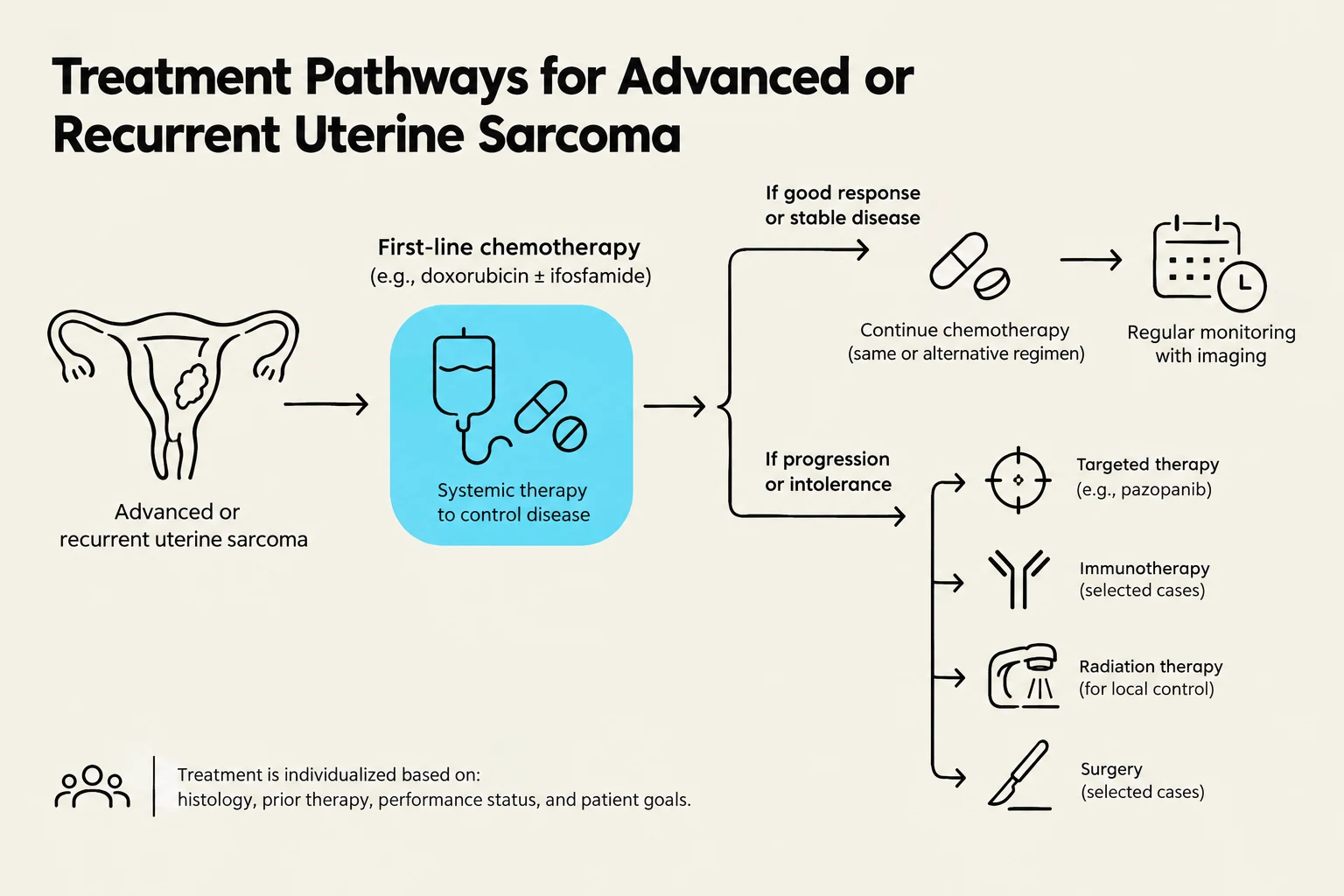
The management of advanced, recurrent, metastatic, or inoperable uterine sarcoma requires a multimodal approach, often involving systemic therapy.
First-Line Therapy for Advanced or Recurrent Disease
For patients with advanced, recurrent, metastatic, or inoperable disease, several systemic chemotherapy regimens are considered first-line options:
Doxorubicin
Docetaxel in combination with Gemcitabine
Doxorubicin in combination with Trabectedin (particularly for leiomyosarcoma)
Doxorubicin in combination with Ifosfamide
Dacarbazine in combination with Doxorubicin
Biomarker-Directed Therapy
The advent of molecular profiling has opened doors for targeted therapies. For tumors identified as positive for NTRK gene fusions, specific inhibitors such as Larotrectinib or Entrectinib are highly effective.
Worked Example: A patient presents with recurrent uterine leiomyosarcoma. Imaging reveals metastatic disease to the lungs. Molecular testing of the tumor tissue reveals an NTRK gene fusion. The treatment team decides to initiate therapy with Larotrectinib, targeting this specific molecular alteration.
Clinical Trials
Given the rarity and complexity of uterine sarcomas, participation in clinical trials is strongly encouraged for patients with advanced, recurrent, or metastatic disease. Clinical trials offer access to novel therapeutic agents and treatment strategies that may improve outcomes.
This diagram illustrates treatment pathways for advanced or recurrent uterine sarcoma, outlining first-line chemotherapy and subsequent options based on patient response, including targeted therapy, immunotherapy, radiation therapy, and surgery.
In conclusion, the management of uterine sarcoma is a complex endeavor requiring a multidisciplinary team approach. Accurate pathological diagnosis, informed by molecular findings, coupled with strategic imaging for staging and surveillance, and tailored systemic therapy for advanced disease, are paramount to optimizing patient outcomes. Continued research into the molecular underpinnings of these rare tumors will undoubtedly lead to further advancements in targeted therapies and personalized treatment strategies.
Keep reading for free.
Create an account to finish this reading and make it part of your study routine.