


The somatosensory system is a complex network that allows the brain to organize, interpret, and use sensory information to create an internal representation of the environment. While we do not strictly require sensory information to move, this feedback is essential for efficient movement and balance. This article explores the specialized receptors that detect position, movement, temperature, and pain, as well as the neural pathways that convey this information to the central nervous system.
Proprioception and Kinesthesia
Proprioception is a comprehensive ability that allows an individual to sense muscle tension, limb or body position, the direction and velocity of movement, muscle effort, and balance. When we specifically sense the movement of our limbs, we are experiencing kinesthesia.
To achieve this, the body relies on specialized mechanoreceptors located within joint capsules, ligaments, and muscles. Key proprioceptors include:
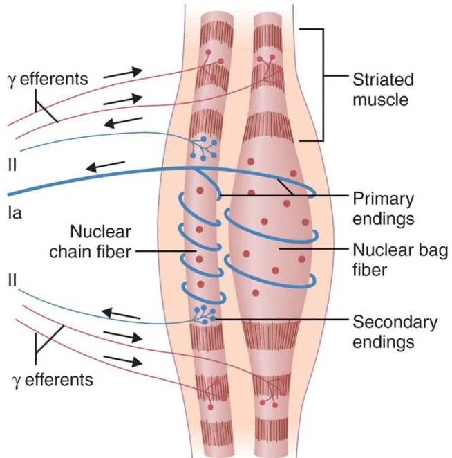
- Muscle Spindles: Located within skeletal muscle, these respond to changes in muscle length.
- Golgi Tendon Organs: Located near the musculotendinous junction, these respond to muscle tension.
- Ruffini Endings: These are activated by skin stretch and contribute to kinesthesia during limb and trunk movement.
Clinical Note: Proprioception is vital for motor control. Impairments in position sense, such as an inability to correctly perceive the position of the great toe, can be a sign of neurological dysfunction.
The Anatomy of the Muscle Spindle
The muscle spindle is a sophisticated sensory organ embedded within the muscle that consists of sensory endings, motor endings, and specialized muscle fibers. It is important to distinguish between the two types of muscle fibers present in a muscle unit:
Keep reading for free.
Create an account to finish this reading and make it part of your study routine.
Continue reading freeAlready have an account? Log in
Updated