Understanding the Human Heart: Anatomy, Function, and Disorders
This comprehensive guide explains the anatomy and function of the human heart, including its role in the cardiovascular system, blood circulation, and electrical conduction. It also details common cardiovascular disorders such as coronary artery disease, arrhythmias, and heart failure.
The human heart is a remarkable muscular organ, roughly the size of a fist, that serves as the central engine of the cardiovascular system. Functioning as a double circulation pump, it ensures that oxygenated blood reaches every cell in the body while simultaneously sending deoxygenated blood to the lungs for gas exchange. Understanding how this pump maintains rhythm, pressure, and flow is essential to understanding how various pathologies—ranging from simple arrhythmias to life-threatening heart failure—arise.
Heart Anatomy and the Double Circulation System
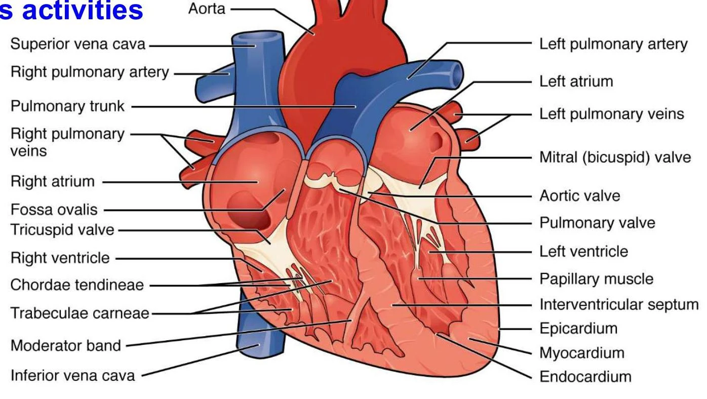
The heart is located within the mediastinum, positioned between the second rib and the fifth intercostal space. It is enclosed in a protective sac called the pericardium and consists of three layers: the epicardium, myocardium (the muscular layer), and endocardium.
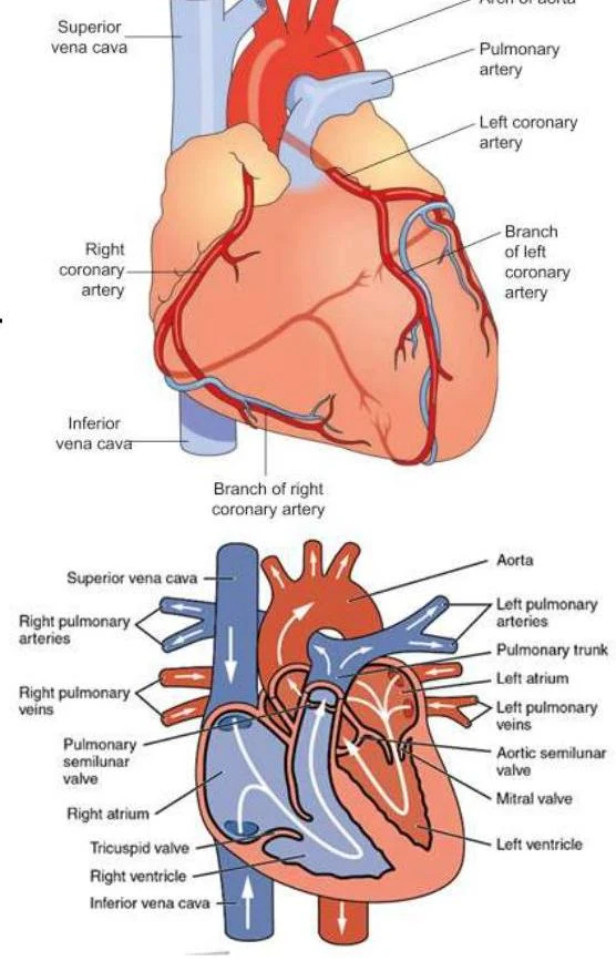
The heart is divided into four chambers: the right and left atria (receiving chambers) and the right and left ventricles (pumping chambers). Because the heart operates a double circulation system, blood passes through it twice in one complete circuit:
Pulmonary Circulation: The right side of the heart receives deoxygenated blood from the body and pumps it through the low-pressure pulmonary circuit to the lungs.
Systemic Circulation: The left side receives oxygenated blood from the lungs and pumps it through the high-pressure systemic circuit to the rest of the body.
Diagram illustrating the anatomy of the human heart, showing its chambers, valves, and major blood vessels including the aorta, pulmonary artery, and vena cava.Diagram of the human heart with various parts labeled, including chambers, valves, and major blood vessels.
A key anatomical distinction is the thickness of the muscular walls. The left ventricle possesses a significantly thicker muscular wall than the right ventricle because it must generate enough force to overcome the high systemic resistance required to pump blood to the entire body.
Cardiac Output and Blood Pressure Regulation
To meet the metabolic demands of the body, the heart must maintain an adequate volume of blood flow. This is measured as Cardiac Output (CO), which is the volume of blood pumped by each ventricle per minute. It is calculated using the following formula:
Cardiac Output=Heart Rate×Stroke Volume
Blood pressure (BP) is the force exerted by the blood against the arterial walls. A normal reading is approximately 120/80 mm Hg. The body regulates BP by adjusting cardiac output and through endocrine mechanisms. When the heart walls are stretched due to high blood volume or high pressure, the heart releases specialized peptides to restore balance:
Atrial Natriuretic Peptide (ANP): Released by the atria.
Brain Natriuretic Peptide (BNP): Produced in the ventricles.
Both peptides act to lower blood pressure by increasing the excretion of sodium (Na+) and water in the kidneys and by inhibiting the Renin-Angiotensin-Aldosterone System (RAAS).
Exam Tip: If a question asks how the heart responds to increased blood volume, remember the "Stretch → Release → Salt/Water Out" logic. Increased stretch triggers ANP/BNP, which promotes sodium and water loss to reduce volume and pressure.
Unidirectional Flow: The Valve System
To ensure that blood moves in only one direction, the heart utilizes four specialized valves. These valves open and close based on pressure differences, and the "lub-dub" sound heard during auscultation is caused by their closing.
Atrioventricular (AV) Valves: Located between the atria and ventricles.
Tricuspid Valve: Between the right atrium and right ventricle.
Bicuspid (Mitral) Valve: Between the left atrium and left ventricle.
Semilunar Valves: Located at the exits of the ventricles.
Pulmonary Valve: Between the right ventricle and the pulmonary artery.
Aortic Valve: Between the left ventricle and the aorta.
Electrical Conduction and the Cardiac Cycle
The heart is myogenic, meaning it exhibits auto-rhythmicity; it generates its own electrical impulses to initiate contraction. The cardiac cycle is the sequence of atrial and ventricular contraction (systole) and relaxation (diastole) that occurs in a single heartbeat.
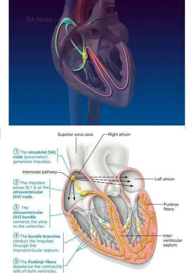
The electrical impulse follows a specific pathway to ensure coordinated contraction:
Sinoatrial (SA) Node: The primary pacemaker, located in the right atrium, which sets the normal sinus rhythm (60–100 beats/min).
Atrioventricular (AV) Node: Receives the impulse from the SA node. There is a slight delay here to allow the ventricles to fill with blood before they contract.
Atrioventricular Bundle (Bundle of His): Carries the impulse toward the ventricles.
Bundle Branches: Distributes the impulse through the interventricular septum.
Purkinje Fibers: Rapidly distribute the impulse throughout the ventricular myocardium, triggering contraction.
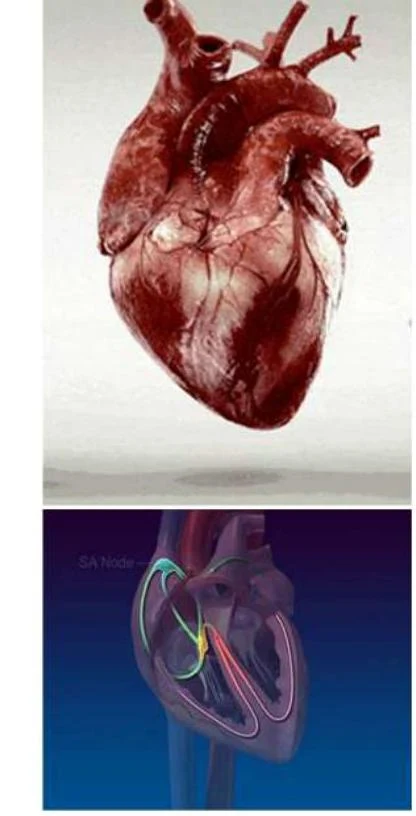
Diagram illustrating the electrical conduction system of the heart, including the SA node, AV node, bundle of His, bundle branches, and Purkinje fibers.An illustration of the human heart, highlighting the SA node, which is responsible for initiating the electrical impulse that causes the heart to beat.
Coronary Circulation: Feeding the Heart
While the heart pumps blood to the entire body, it requires its own dedicated blood supply to power the myocardium. This is provided by the coronary arteries, which arise from the base of the aorta:
Left Coronary Artery: Branches into the left circumflex and the left anterior descending (interventricular) arteries. It supplies the left atrium and much of the left ventricle.
Right Coronary Artery: Supplies the right atrium and most of the right ventricle.
Diagrams illustrating the coronary arteries and semilunar valves of the heart.Anatomical illustration of the human heart, showing anterior and posterior views with labeled coronary arteries and veins.
Deoxygenated blood from the myocardium is collected by cardiac veins, which merge into the coronary sinus, eventually emptying into the right atrium. Notably, coronary blood flow to the left ventricle is greatest during diastole (relaxation), as the muscular contraction during systole can compress the vessels.
Cardiovascular Disorders
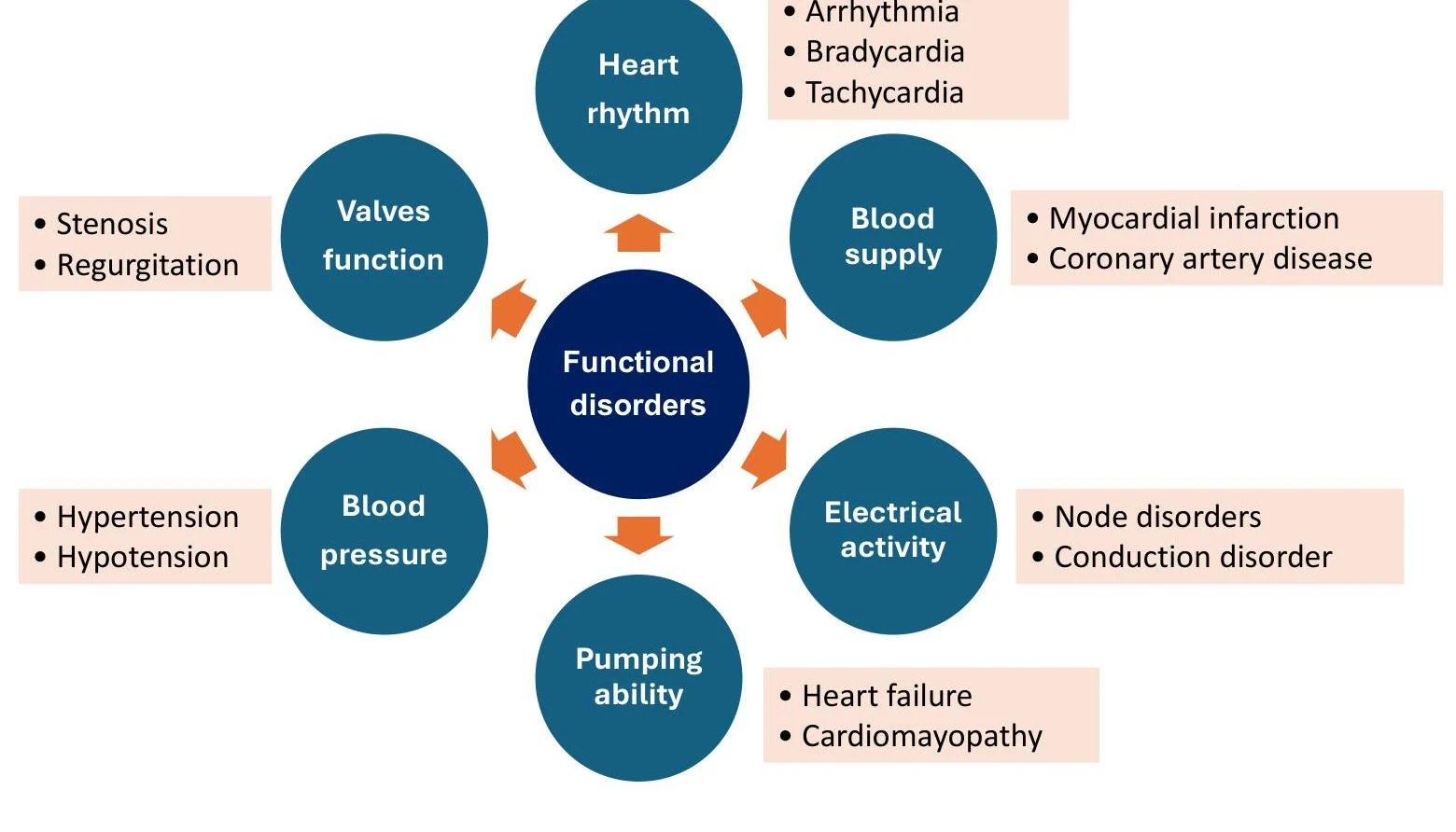
Cardiovascular disorders can be categorized by the specific component of the system that fails: rhythm/conduction, coronary blood supply, valve function, pumping strength, or pressure regulation.
A primary driver of many of these issues is atherosclerosis, the buildup of plaque within the arterial walls.
Common Pathologies:
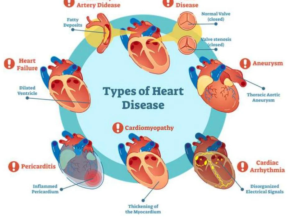
Coronary Artery Disease (CAD): Occurs when the coronary arteries become narrowed or blocked by plaque, reducing oxygen delivery to the heart muscle. This can manifest as angina (chest pain) or lead to a myocardial infarction (heart attack).
Cardiac Arrhythmia: A failure in the electrical conduction system, resulting in an irregular heartbeat. For example, if the SA node fails, the AV node may take over as an "escape pacemaker," but at a much slower rate (around 45–50 beats/min).
Heart Failure: A condition where the heart cannot pump blood effectively enough to meet the body's needs. This can be categorized into systolic failure (pumping strength) or diastolic failure (filling/pressure issues).
Valvular Disorders: Problems with the opening or closing of the heart valves.
This diagram illustrates various functional disorders of the heart, categorized by heart rhythm, valves function, blood supply, electrical activity, pumping ability, and blood pressure.This diagram illustrates various types of heart disease, including artery disease, heart failure, pericarditis, cardiomyopathy, aneurysm, and cardiac arrhythmia.
Clinical Correlation: A patient presenting with dizziness and an ECG showing a regular heart rate of 45 bpm without P waves is likely experiencing a junctional rhythm, where the AV node has taken over as the pacemaker because the SA node is not functioning.
Keep reading for free.
Create an account to finish this reading and make it part of your study routine.