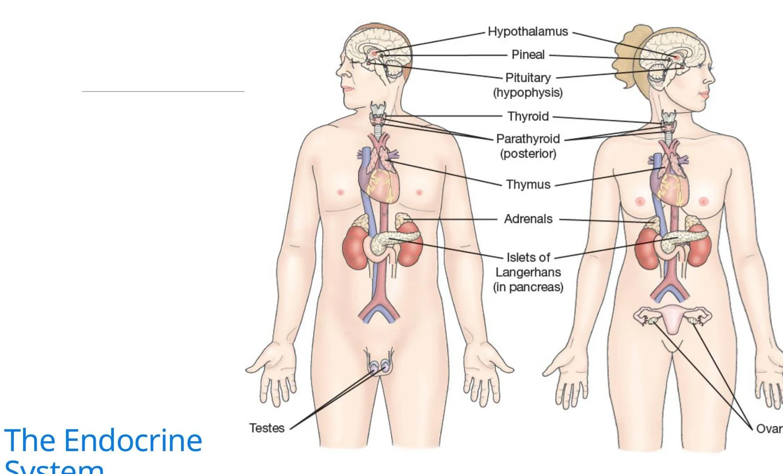
- ACTH (Adrenocorticotropic hormone): Stimulates the adrenal cortex to release aldosterone and cortisol.
- FSH (Follicle Stimulating Hormone) & LH (Luteinizing Hormone): Regulate reproductive functions in both men and women.
- GH (Growth Hormone): Promotes growth, development, and anabolic metabolism.
- PRL (Prolactin): Stimulates milk production in the breasts.
- TSH (Thyroid Stimulating Hormone): Stimulates the thyroid gland to release T3 and T4.
The Posterior Pituitary
Unlike the anterior lobe, the posterior pituitary does not produce its own hormones; instead, it stores and secretes hormones produced by the hypothalamus:
- ADH (Antidiuretic Hormone): Signals the kidneys to reabsorb water, increasing blood volume and decreasing blood pressure.
- Oxytocin: Stimulates uterine contractions during labor and milk ejection during breastfeeding.
Clinical Alert: Pituitary Pathophysiology
Abnormalities in the pituitary often stem from over- or undersecretion. Hypopituitarism (undersecretion) can result from disease, trauma, or radiation and may lead to extreme weight loss, hair loss, impotence, and even coma or death if hormones are not replaced.
Specific imbalances in ADH secretion lead to two critical disorders: Diabetes Insipidus (deficiency of ADH) and SIADH (Syndrome of Inappropriate Antidiuretic Hormone, or excess ADH).
Nursing Assessment and Diagnostics
Effective management begins with a thorough clinical assessment to identify metabolic and fluid imbalances.
Health History and Physical Examination
Nurses should prioritize asking about changes in energy levels, weight, thirst, urination, bowel function, and mood. Physical assessment should focus on:
- Vital Signs: Monitoring for changes in heart rate and blood pressure.
- Skin and Hair: Checking for thinning skin, irregular hair growth, or changes in texture.
- Physical Landmarks: Looking for exophthalmos (protruding eyeballs), moon face, buffalo hump (fat pad on the upper back), and edema.
Diagnostic Studies
- Blood Tests: Used to measure circulating hormone levels (e.g., T3, T4, and TSH) and autoantibodies.
- Urine Tests: Often involve 24-hour collections to measure hormone excretion, such as catecholamines.
- Stimulation vs. Suppression Tests: Stimulation tests are used to confirm hypofunction (the gland's response to a stimulus), while suppression tests are used to detect hyperfunction (the effect of an exogenous hormone dose).
- Imaging: MRI, CT, ultrasound, and radioactive scans help visualize gland size, shape, and function.
Major Endocrine Disorders
Thyroid Disorders: "Low and Slow" vs. "Amped Up"
The thyroid gland produces T4 (thyroxine), T3 (triiodothyronine), and calcitonin.
1. Hypothyroidism ("Low and Slow"):
Characterized by low T3 and T4 levels. Patients often present with fatigue, weight gain, cold intolerance, bradycardia, and depression. A life-threatening complication is Myxedema coma, which involves cardiovascular collapse and unconsciousness.
- Management: Lifelong hormone replacement therapy (e.g., Levothyroxine). Patients must take this before breakfast on an empty stomach.
2. Hyperthyroidism ("Amped Up"):
Characterized by excessive hormone secretion, often due to Graves' disease. Symptoms include anxiety, tachycardia, weight loss, heat intolerance, and exophthalmos. The most severe complication is Thyroid Storm, a hypermetabolic crisis characterized by high fever (>38.5∘C), extreme tachycardia, and delirium.
- Management: Antithyroid medications (Propylthiouracil, Methimazole) and beta-blockers to control cardiac symptoms.
Parathyroid Disorders: Calcium Regulation
The parathyroid glands secrete Parathyroid Hormone (PTH), which increases blood calcium levels.
- Hypoparathyroidism: Results in low calcium (hypocalcemia) and high phosphorus. This can cause tetany (muscle spasms), numbness, and tingling. Nurses must monitor for Chvostek's or Trousseau's signs.
- Hyperparathyroidism: Results in high calcium (hypercalcemia), which can lead to bone decalcification, kidney stones (nephrolithiasis), and abdominal pain.
Adrenal Disorders: Stress and Metabolism
The adrenal glands consist of the medulla (secretes catecholamines like epinephrine) and the cortex (secretes steroids like cortisol and aldosterone).
1. Addison’s Disease (Adrenal Insufficiency):
An "absence of steroids". Patients experience low blood pressure, low glucose, low sodium, and high potassium. They often crave salt and may show bronze skin pigmentation.
- Management: Lifelong steroid replacement (e.g., Prednisone) and high-sodium/high-protein diets.
2. Cushing’s Syndrome (Adrenal Excess):
Characterized by excessive cortisol. Clinical signs include truncal obesity, "moon face," "buffalo hump," hypertension, hyperglycemia, and thin skin with stretch marks.
- Management: Controlling the underlying cause (such as tumor removal) and managing complications like diabetes and osteoporosis.
Nursing Tip: Medication Safety
When managing patients on steroids (for either Addison's or Cushing's), never advise them to stop the medication abruptly, as this can trigger a life-threatening crisis.