


To understand how the heart functions, one must look beyond the physical muscle and into the electrical signals that command it. An Electrocardiogram (ECG or EKG) is a visual representation of these electrical impulses as they travel through the heart muscle. By studying the characteristic "PQRST" waveform, clinicians and students can interpret the timing and health of the heart's conduction system.
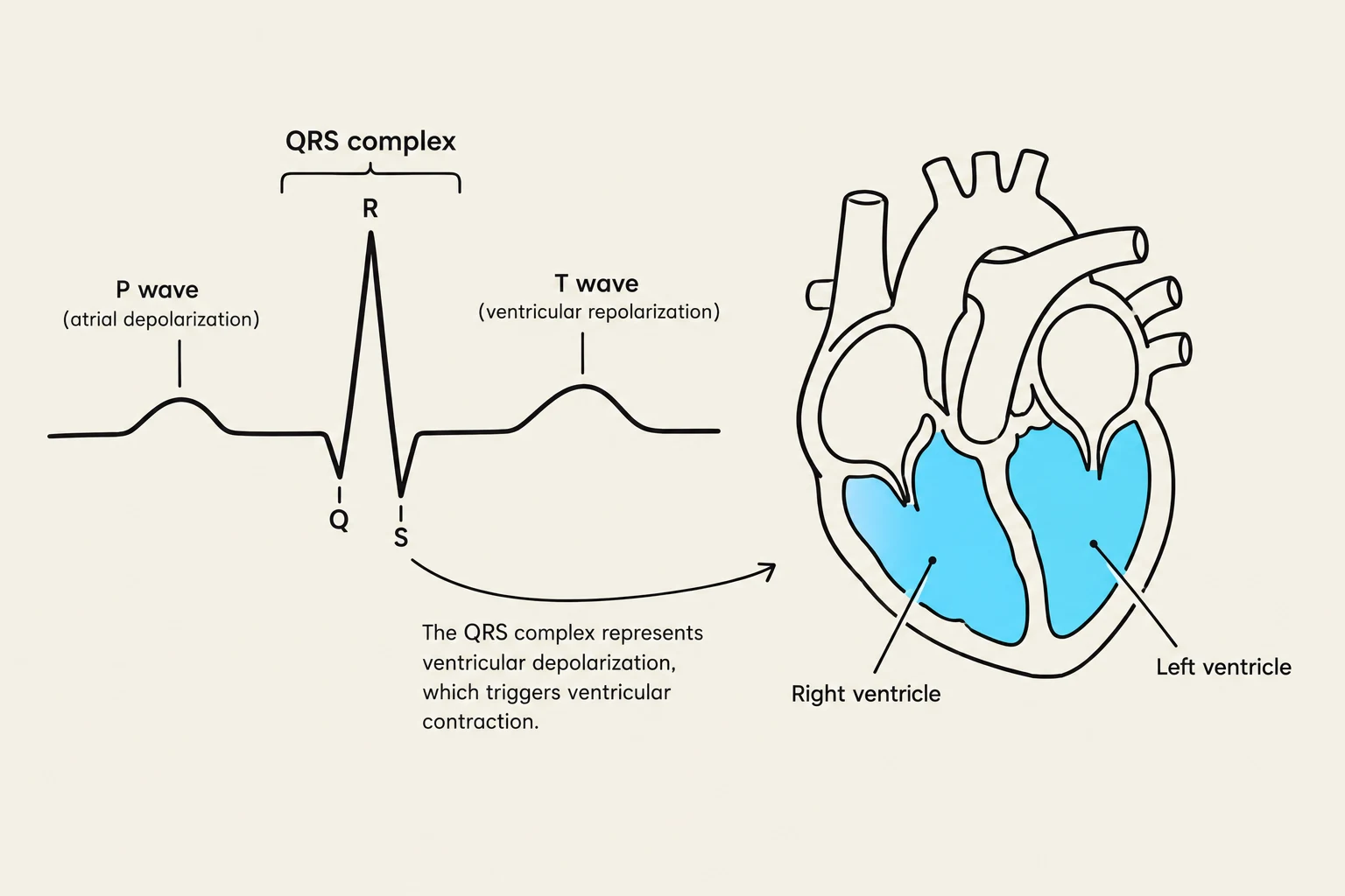
This article breaks down the cardiac cycle into its four primary electrical components: the P-wave, the PR segment, the QRS complex, and the T-wave, linking each electrical event to its corresponding mechanical action in the heart.
1. The P-Wave: Atrial Activation
The cardiac cycle begins with a spontaneous electrical impulse generated by the SA node (Sinoatrial node), often referred to as the heart's natural pacemaker. This impulse triggers atrial depolarization, which is the electrical process that prepares the upper chambers of the heart to contract.
On an ECG tracing, this is seen as a small, rounded upward deflection known as the P-wave. Anatomically, during this phase, the atria (the upper chambers) become active and begin to contract to push blood into the ventricles.

Exam Tip: If you see an absent or irregular P-wave on an ECG, it often indicates that the SA node is not properly initiating the heartbeat, potentially pointing toward an arrhythmia.
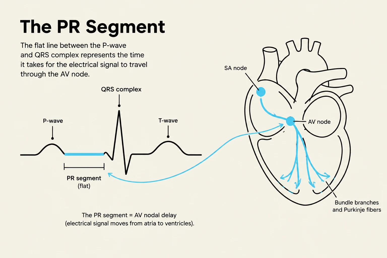
2. The PR Segment: The Controlled Delay
Once the atria have depolarized, the electrical impulse reaches the AV node (Atrioventricular node). This leads to the PR segment, which appears as a flat period on the ECG tracing between the end of the P-wave and the start of the QRS complex.
Keep reading for free.
Create an account to finish this reading and make it part of your study routine.
Continue reading freeAlready have an account? Log in
Updated