An overview of the autonomic nervous system, its divisions (sympathetic and parasympathetic), neural anatomy, neurotransmitters, receptors, and physiological effects on various organs. It also touches upon autonomic reflex arcs.
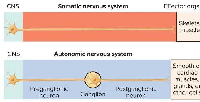
The autonomic nervous system (ANS) is a vital component of the peripheral nervous system responsible for the involuntary control of most tissue and organ systems. Unlike the somatic nervous system, which coordinates the voluntary control of skeletal muscles, the ANS regulates visceral effector organs such as smooth muscle, cardiac muscle, glands, and even adipocytes (fat cells). This system ensures that your body maintains homeostasis by automatically adjusting physiological processes without the need for conscious thought.
Divisions of the Autonomic Nervous System
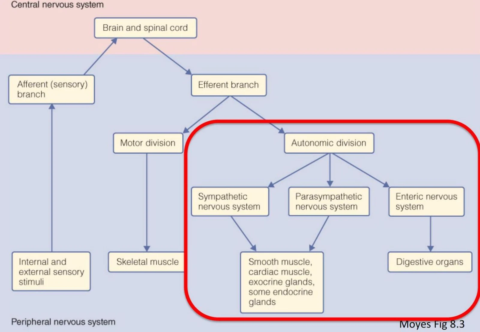
The ANS is functionally divided into two primary branches that typically act in opposition to one another: the sympathetic division and the parasympathetic division. This relationship is often compared to a car with one foot on the accelerator and the other on the brake; as one division increases its activity, the other typically decreases.
Sympathetic Division: Known as the "fight-or-flight" system, this division is most active during periods of stress or physical activity. It prepares the body for intense physical exertion by increasing heart rate, dilating pupils, and mobilizing energy stores.
Parasympathetic Division: Known as the "rest-and-digest" system, this division is most active during periods of rest. It focuses on energy conservation, digestion, and other restorative processes.
A third, specialized component is the enteric nervous system, which is located within the walls of the gastrointestinal tract. While often classified as a subdivision of the autonomic efferent system, it is unique because it also contains its own sensory neurons and interneurons.
Diagram illustrating the divisions of the nervous system, including the central and peripheral nervous systems, and further subdivisions like the autonomic, sympathetic, parasympathetic, and enteric nervous systems.
Neural Anatomy: Preganglionic and Postganglionic Neurons
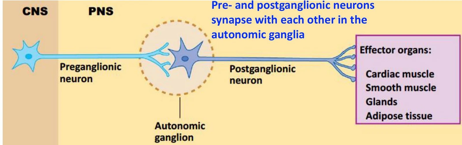
The structural hallmark of the autonomic nervous system is that it utilizes a two-neuron chain to connect the central nervous system (CNS) to the target organ.
This diagram illustrates the somatic and autonomic nervous systems, showing their respective pathways from the central nervous system (CNS) to effector organs like skeletal muscles or smooth/cardiac muscles and glands.
Preganglionic Neurons: These neurons have their cell bodies located within the brainstem or spinal cord. Their axons project outward to meet the second neuron in the chain.
Autonomic Ganglia: The synapse between the preganglionic and postganglionic neurons occurs outside the CNS in a cluster of nerve cells called an autonomic ganglion.
Postganglionic Neurons: The cell bodies of these neurons reside within the autonomic ganglia, and their axons project from the ganglia to the final visceral effector organ.
Diagram illustrating the connection between the central nervous system (CNS) and peripheral nervous system (PNS) through preganglionic and postganglionic neurons synapsing in autonomic ganglia, which then innervate effector organs like cardiac muscle, smooth muscle, glands, and adipose tissue.
Anatomical Differences Between Divisions
The two divisions differ significantly in where they exit the CNS and where their ganglia are located:
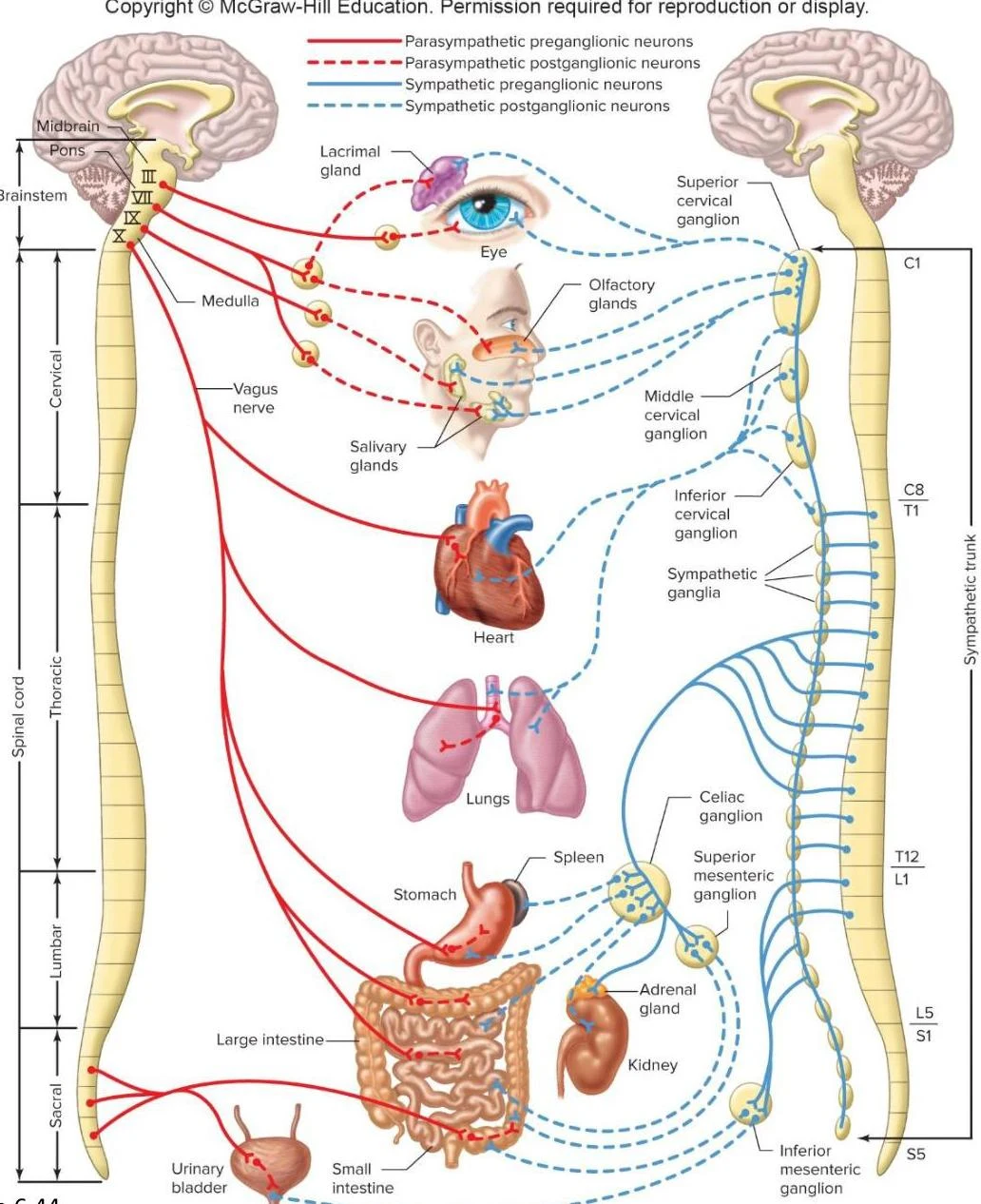
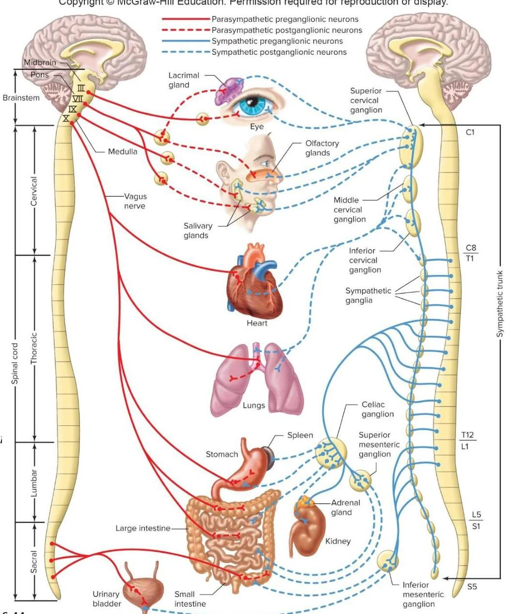
Sympathetic (Thoracolumbar) Division: These neurons exit the spinal cord from the thoracic and lumbar regions. Most sympathetic ganglia are located close to the spinal cord in structures called sympathetic trunks (or paravertebral ganglia). Some sympathetic fibers also synapse in collateral ganglia located in the abdominal cavity. Consequently, sympathetic preganglionic neurons are relatively short, while postganglionic neurons are long.
Parasympathetic (Craniosacral) Division: These neurons exit from the brainstem and the sacral portion of the spinal cord. Their ganglia are located very near or even within the walls of the target organs, resulting in long preganglionic neurons and short postganglionic neurons.
Diagram illustrating the human autonomic nervous system, detailing the pathways of the parasympathetic and sympathetic nervous systems and their connections to various organs.
Neurotransmitters and Receptors
The communication within the ANS relies on specific chemical messengers called neurotransmitters and the specialized proteins on target cells called receptors.
The Cholinergic System (Acetylcholine)
Acetylcholine (ACh) is used by all preganglionic neurons in both the sympathetic and parasympathetic divisions.
In the parasympathetic division, ACh is also the neurotransmitter used by the postganglionic neuron to signal the effector organ.
The receptors on the postganglionic neurons in the ganglia are nicotinic receptors (ionotropic).
The receptors on the target tissues of the parasympathetic system are muscarinic receptors (metabotropic).
The Adrenergic System (Norepinephrine)
In the sympathetic division, the postganglionic neuron usually releases norepinephrine (NE) onto the target tissue.
The receptors on the target tissues for norepinephrine are known as adrenergic receptors, which are divided into two main subtypes: alpha (α) and beta (β).
Note: A small exception exists where some sympathetic postganglionic fibers release ACh (e.g., to sweat glands).
The Adrenal Medulla
A unique component of the sympathetic system is the adrenal medulla. Instead of having long axons, a set of sympathetic postganglionic neurons forms part of this endocrine gland. When activated, the adrenal medulla releases epinephrine (about $80%$) and norepinephrine (about $20%$) directly into the bloodstream to act as hormones.
Exam Tip: Remember the "Two-A" rule for the Sympathetic Postganglionic side: Adrenaline (Epinephrine/Norepinephrine) and Adrenergic receptors (α and β). For the Parasympathetic side, think Acetylcholine and Muscarinic receptors.
Dual Innervation and Physiological Effects
Most visceral organs receive dual innervation, meaning they are supplied by both sympathetic and parasympathetic fibers. This allows for precise, reciprocal control over organ function.
Diagram illustrating the autonomic nervous system's control over various organs, differentiating between parasympathetic and sympathetic pathways.
The following table summarizes how these divisions affect various organs:
Effector Organ
Sympathetic Effect (Receptor)
Parasympathetic Effect (Muscarinic)
Eyes (Iris)
Dilates pupil (α1)
Constricts pupil
Heart (SA Node)
Increases heart rate (β1)
Decreases heart rate
Lungs (Bronchi)
Relaxes/Dilates (β2)
Contracts
Stomach/Intestine
Decreases motility/secretion (α,β)
Increases motility/secretion
Bladder Wall
Relaxes (β2)
Contracts
Autonomic Reflex Arcs
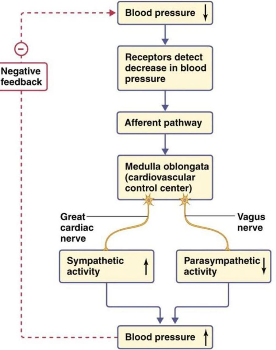
Many autonomic responses are not controlled by conscious thought but instead occur through autonomic reflex arcs. These are simple neural circuits that allow the body to respond to changes (such as blood pressure fluctuations) via negative feedback loops to maintain stability without brain intervention.
This diagram illustrates the baroreceptor reflex, a negative feedback mechanism that regulates blood pressure. When blood pressure drops, receptors detect this change, sending signals via the afferent pathway to the medulla oblongata. The cardiovascular control center then adjusts sympathetic and parasympathetic activity to restore blood pressure.
Keep reading for free.
Create an account to finish this reading and make it part of your study routine.