Understanding the Auditory System: Cochlea, Stereocilia, and Eustachian Tube
An explanation of the human auditory system, detailing the functions of the Eustachian tube for pressure equalization, the cochlea's role in converting vibrations to nerve impulses, and the microscopic process of stereocilia in auditory transduction.
The human auditory system is a complex network of structures designed to capture sound waves, convert them into mechanical vibrations, and ultimately transform those vibrations into electrical impulses that the brain can interpret. While we often think of hearing as a single event, it is actually a multi-stage process involving the external, middle, and inner ear. To understand how we hear—and how we maintain balance—we must look closely at the specialized anatomy of the cochlea, the microscopic function of stereocilia, and the regulatory role of the Eustachian tube.
The Middle Ear and the Eustachian Tube
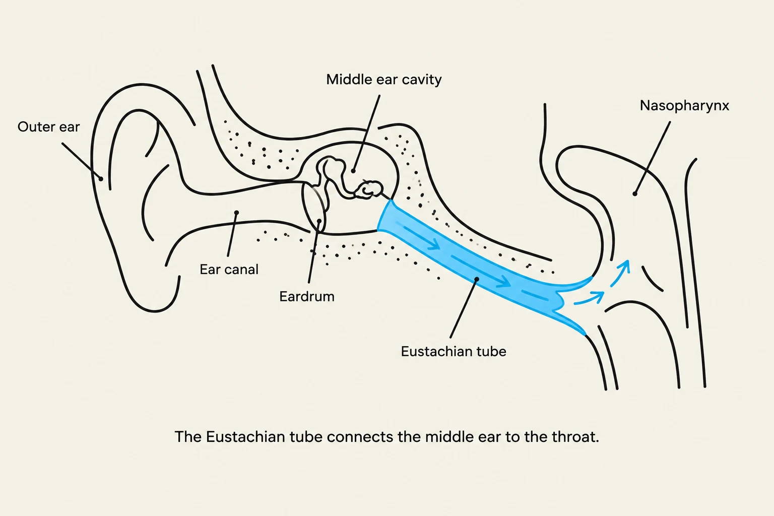
Diagram illustrating the anatomy of the human ear, including the outer ear, ear canal, eardrum, middle ear cavity, Eustachian tube, and nasopharynx. The Eustachian tube is shown connecting the middle ear to the throat.
Before sound can reach the delicate structures of the inner ear, it must pass through the external auditory canal and strike the tympanic membrane (eardrum). The tympanic membrane serves to capture sound waves and begin the process of vibration. These vibrations are then passed to the ossicles—the tiny bones of the middle ear—which intensify the mechanical energy before it enters the fluid-filled inner ear.
A critical component of this middle ear environment is the Eustachian tube. This narrow canal connects the middle ear to the nasopharynx (the upper part of the throat).
The Function of the Eustachian Tube
The primary role of the Eustachian tube is pressure equalization. For the tympanic membrane to vibrate freely and efficiently, the air pressure inside the middle ear must match the atmospheric pressure outside the body.
Pressure Regulation: When you swallow or yawn, the Eustachian tube opens momentarily, allowing air to enter or leave the middle ear to equalize pressure.
Drainage: It also helps drain secretions from the middle ear into the nasopharynx, preventing fluid buildup that could impair hearing.
Exam Tip: If a question asks about the connection between the middle ear and the nasopharynx, the answer is the Eustachian tube. Remember that its primary job is pressure equalization!
The Inner Ear: The Cochlea
The cochlea converts mechanical vibrations into fluid waves.
Once the mechanical vibrations from the ossicles reach the inner ear, they encounter a different medium: fluid. The cochlea is the snail-shaped, fluid-filled structure within the inner ear responsible for the actual process of hearing.
The cochlea acts as a biological transducer. It takes the mechanical energy (vibrations) and converts it into electrochemical energy (nerve impulses). As the vibrations enter the cochlea, they create waves in the internal fluids. These fluid waves travel through the cochlear chambers, eventually triggering the sensory cells that send signals to the brain via the vestibulocochlear nerve.
Microscopic Transduction: Stereocilia
The true "magic" of hearing happens at a microscopic level within the cochlea. Inside the cochlea are specialized sensory cells (hair cells) that possess hair-like projections known as stereocilia.
How Stereocilia Work
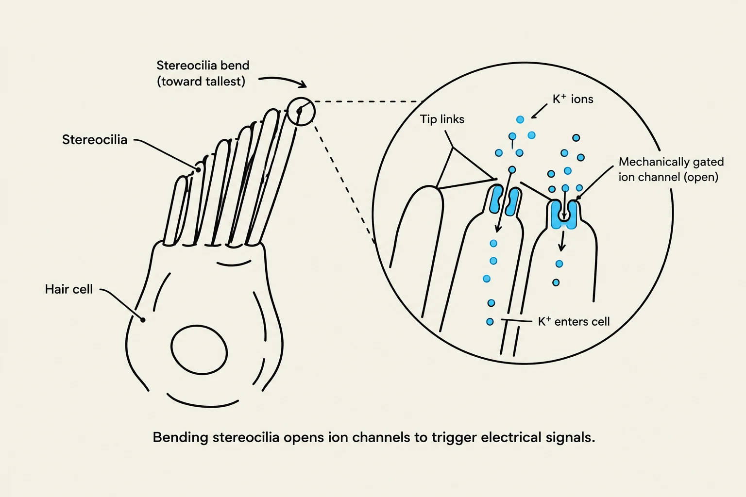
Bending stereocilia opens ion channels to trigger electrical signals.
Stereocilia are the structures responsible for translating fluid movement into electrical signals. The process follows these steps:
Fluid Movement: As sound waves move through the cochlear fluid, the movement causes the stereocilia to bend.
Mechanical Gating: The bending of these stereocilia opens mechanically gated ion channels located at their tips.
Depolarization: The influx of ions (primarily potassium) into the cell causes a change in the electrical charge of the cell, known as depolarization.
Neural Signaling: This electrical change triggers the release of neurotransmitters, which then stimulate the auditory nerve to send a signal to the brain.
Clinical Correlation: The loss of these stereocilia is a significant factor in age-related hearing loss. When these microscopic structures are damaged by loud noises or aging, the ability to convert sound into electrical impulses is diminished.
Summary of Auditory Pathways
To visualize the flow of information, we can summarize the process as follows:
Structure
Medium
Primary Function
Tympanic Membrane
Air → Solid
Captures sound waves and initiates vibration
Eustachian Tube
Air
Equalizes pressure between middle ear and nasopharynx
Cochlea
Fluid
Translates mechanical vibrations into fluid waves
Stereocilia
Cellular/Ionic
Converts fluid movement into electrical impulses via depolarization
By understanding these interconnected parts—from the macro-level pressure regulation of the Eustachian tube to the micro-level ion movement of the stereocilia—we gain a complete picture of how the human body perceives the world of sound.
Keep reading for free.
Create an account to finish this reading and make it part of your study routine.