


In the study of electrocardiography (ECG), understanding sinus rhythms is the fundamental starting point for any clinician. Sinus rhythms represent the heart's natural, healthy electrical conduction pattern. While an ECG provides a graphic display of the heart's electrical activity, it is vital to remember that this electrical impulse is distinct from the heart's mechanical activity—the actual physical contraction of the muscle.
The Origin: The SA Node
All sinus rhythms originate in the Sinoatrial (SA) node. The SA node acts as the heart's natural pacemaker, initiating the electrical impulse that travels through the heart's conduction pathway in a predictable pattern. This impulse triggers atrial depolarization (contraction), followed by the impulse traveling through the AV node to trigger ventricular depolarization (contraction).
Morphological Characteristics of Sinus Rhythms

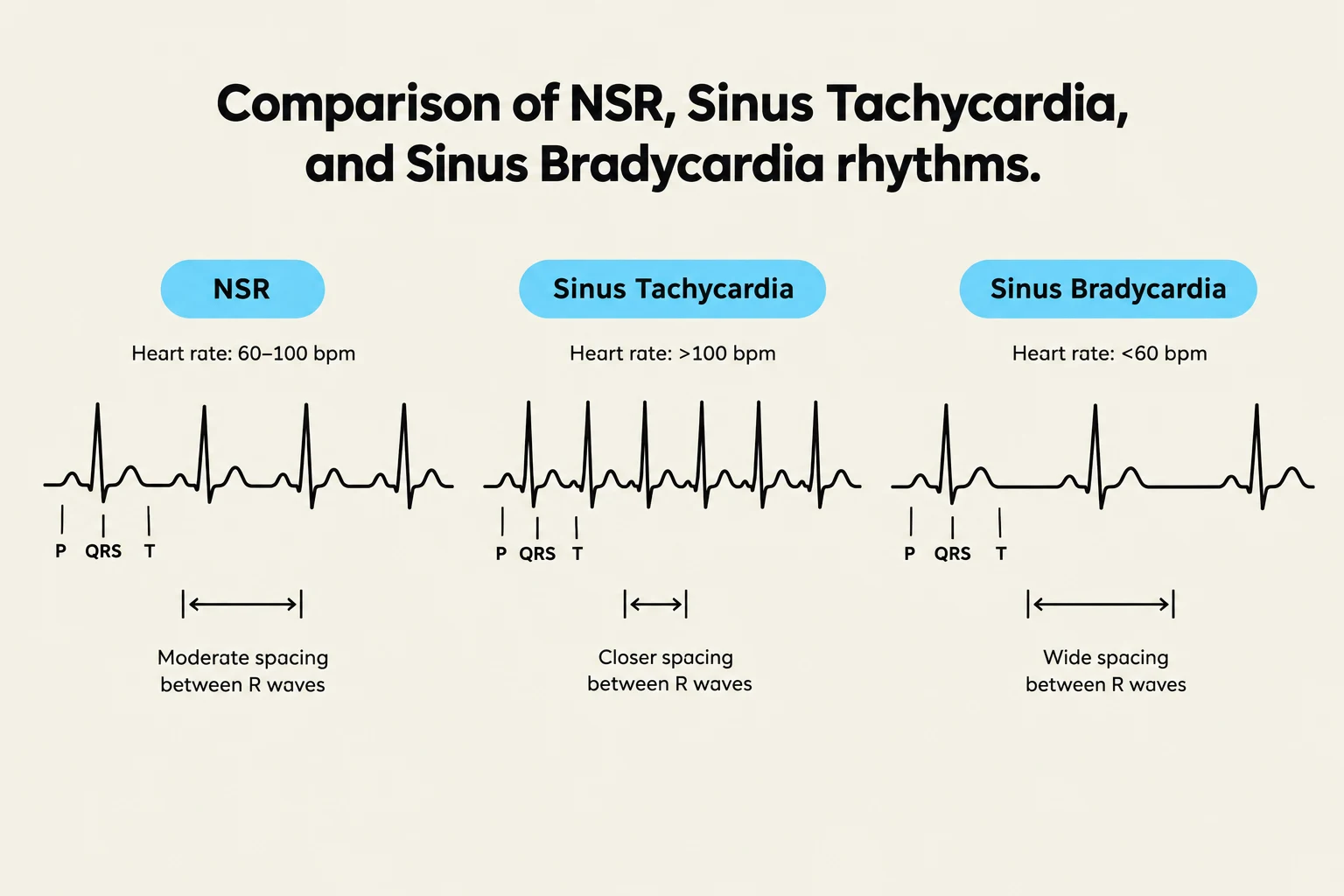
- The P Wave: This wave represents atrial contraction. In a sinus rhythm, the P wave must be symmetrical and upright in Lead II, with exactly one P wave preceding every QRS complex.
- The PR Interval: This measures the time from the start of the P wave to the start of the QRS complex, representing the normal delay at the AV node. A normal PR interval is consistent and measures between and seconds (equivalent to 3–5 tiny boxes on ECG paper).
- The QRS Complex: This represents ventricular contraction. In sinus rhythms, the QRS complex is narrow, measuring between and seconds.
- Rhythm Regularity: The rhythm must be regular, meaning the distance between consecutive R waves is consistent.
Keep reading for free.
Create an account to finish this reading and make it part of your study routine.
Continue reading freeAlready have an account? Log in
Updated