


When assessing a patient on a cardiac monitor, your first priority is never the rhythm strip itself, but the patient. Always begin with a clinical assessment of responsiveness, breathing, pulse, blood pressure, saturation, and signs of perfusion. If a patient is unresponsive and lacks a pulse, you must immediately activate emergency protocols and begin CPR. It is critical to distinguish between shockable rhythms, such as Ventricular Tachycardia (Vt) and pulseless Vt, and non-shockable rhythms, such as Asystole and Pulseless Electrical Activity (PEA).
In this article, we will focus on the "sinus-based" rhythms—those originating from the sinoatrial (SA) node—and the first common deviation from this pattern: the Premature Atrial Contraction (PAC).
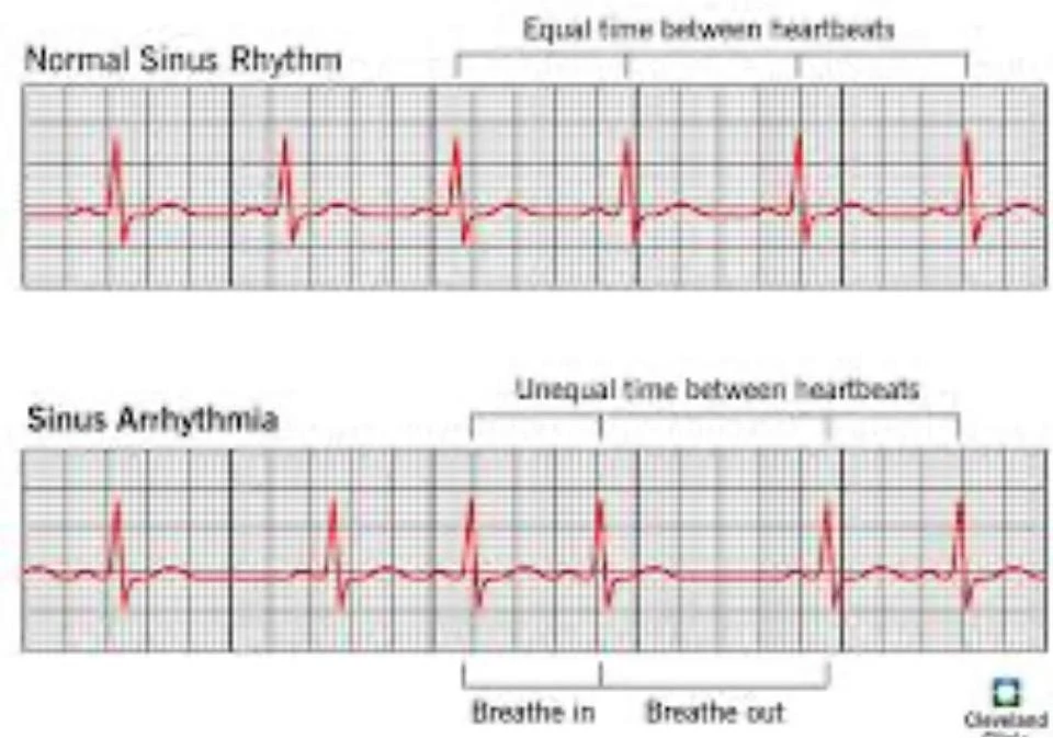
Normal Sinus Rhythm (NSR)
Normal Sinus Rhythm is the baseline for all cardiac rhythm analysis; every other rhythm is compared against this standard. NSR is defined by a regular pattern where the heart rate is between .
The key ECG clues for NSR include:
- A consistent P wave morphology before every QRS complex.
- A regular rhythm with a predictable R-R interval.
Patients in NSR are typically comfortable and demonstrate adequate perfusion. While the rhythm itself is normal, clinicians should always investigate any symptoms a patient reports, even if the ECG strip appears perfect.
Sinus Bradycardia
Sinus bradycardia is a sinus pattern where the rate is below .

While a slow heart rate can be normal in certain populations, such as athletes or during sleep, it becomes clinically significant when the patient becomes symptomatic. Symptoms typically emerge when the rate falls below and may include dizziness, fatigue, fainting, confusion, or hypotension. Common causes include vagal stimulation (such as vomiting or suctioning), medications like beta-blockers, hypoxia, ischemia, or hypothyroidism.


Keep reading for free.
Create an account to finish this reading and make it part of your study routine.
Continue reading freeAlready have an account? Log in
Updated