


In the study of histology, identifying tissues requires a keen eye for both cell shape and the arrangement of those cells. One of the most distinct and functionally specialized tissue types is the simple cuboidal epithelium. While it may not be as complex as the stratified layers found in the skin, its specific structure makes it indispensable for the physiological processes of the body.
Defining the Structure
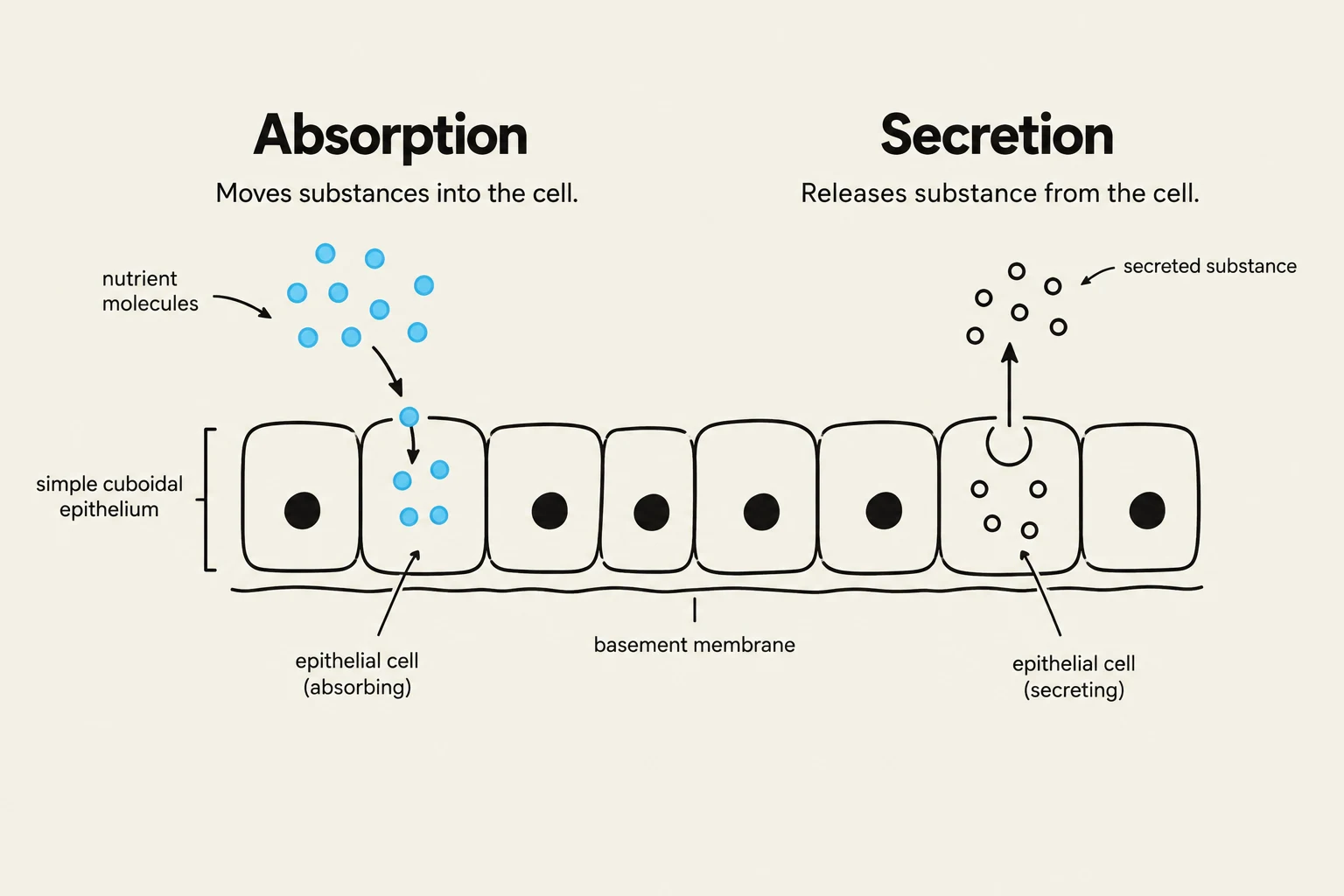
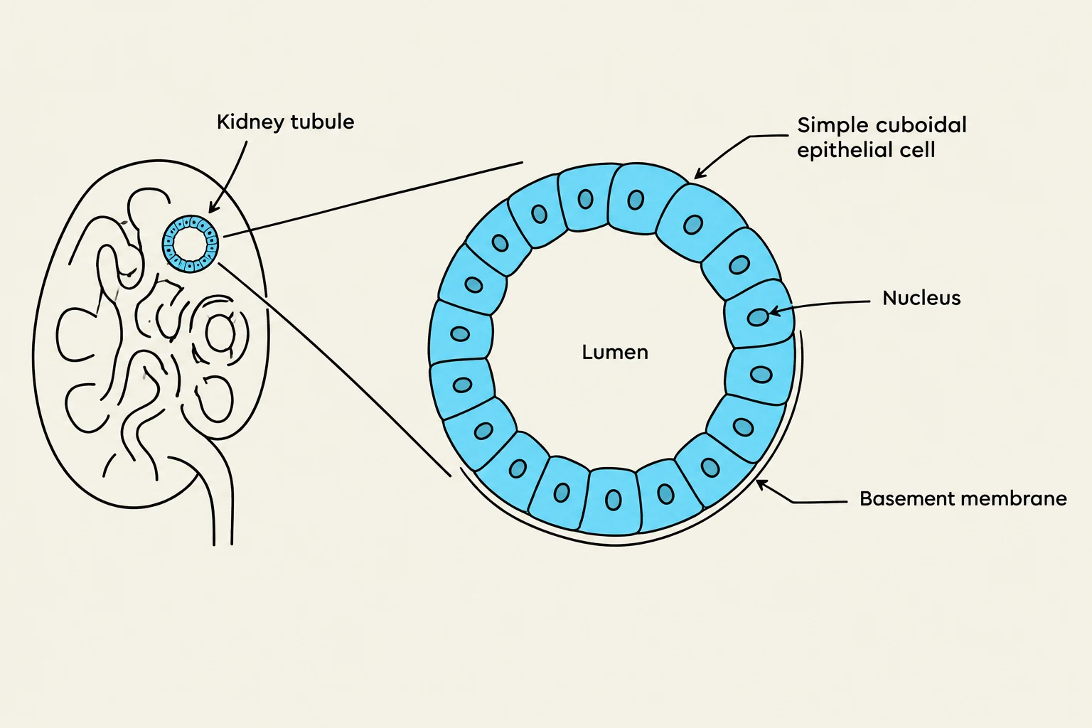
To identify simple cuboidal epithelium, you must look for two primary characteristics: the number of cell layers and the individual shape of the cells. The term "simple" refers to the fact that the tissue consists of a single layer of cells resting on a basement membrane. The term "cuboidal" describes the geometry of the cells themselves.
When viewed under a microscope, these cells appear roughly as square or cube-shaped units. Unlike squamous cells, which are flat and scale-like, or columnar cells, which are tall and narrow, cuboidal cells have a width and height that are approximately equal. A hallmark of this tissue type is the position of the nucleus; because the cell is roughly equal in all dimensions, the nucleus is typically large, spherical, and located centrally within the cell.

Exam Tip: When looking at a slide, if you see a single row of cells where the nucleus looks like a perfect circle in the middle of a square-ish cell, you are likely looking at simple cuboidal epithelium.
Keep reading for free.
Create an account to finish this reading and make it part of your study routine.
Continue reading freeAlready have an account? Log in
Updated