Understanding Shock: Types, Pathophysiology, and Complications
An in-depth explanation of shock, a critical medical emergency characterized by inadequate tissue perfusion. This guide covers the cellular crisis of hypoxia, the body's compensatory mechanisms, and the four primary classifications of shock: cardiogenic, hypovolemic, obstructive, and distributive (including septic, ana
Shock is not merely "low blood pressure"; it is a critical, life-threatening medical emergency defined by inadequate perfusion to the body's tissues. When the delivery of oxygen and nutrients to cells fails to meet their metabolic demands, a cascade of cellular dysfunction begins. This failure can stem from a lack of blood volume, a failure of the heart to pump, a physical blockage in the vessels, or a massive loss of vascular tone. Understanding the specific type of shock is vital, as the treatment for one type (such as aggressive fluid resuscitation) could be detrimental to another (such as cardiogenic shock).
The Cellular Crisis: Hypoxia and Compensation
At the heart of all shock states is the transition from aerobic to anaerobic metabolism. When perfusion drops, cells cannot receive enough oxygen, leading to cellular hypoxia. Without oxygen, cells cannot produce ATP efficiently, leading to cellular injury and, eventually, cell death.
To combat this, the body initiates powerful compensatory mechanisms to maintain blood pressure and perfusion to vital organs like the brain and heart.
Sympathetic Nervous System (SNS) Activation: The body releases catecholamines (like adrenaline), which increase heart rate (HR) and cause vasoconstriction to redirect blood to the core.
Renin-Angiotensin-Aldosterone System (RAAS) Activation: The kidneys detect decreased perfusion and release renin. This leads to the production of Angiotensin II, a potent vasoconstrictor, and stimulates the release of aldosterone, which causes the kidneys to retain sodium and water to increase blood volume.
Exam Tip: While blood pressure is a common vital sign, it is not always an accurate indicator of early shock. A patient may maintain a "normal" BP through intense compensatory vasoconstriction even while they are slipping into profound shock.
The Four Primary Classifications of Shock
Shock is categorized based on the underlying physiological mechanism causing the perfusion failure.
1. Cardiogenic Shock: "The Pump Fails"
In cardiogenic shock, the heart is unable to maintain adequate cardiac output despite having enough blood volume. This is often caused by a myocardial infarction (heart attack), cardiomyopathies, or cardiac trauma.
Clinical Manifestations:
Hypotension
Cyanotic (bluish) nail beds and mucosa, indicating poor oxygenation
Decreased urine output due to poor renal perfusion
Altered level of consciousness (LOC)
2. Hypovolemic Shock: "Not Enough Fluid"
This occurs when there is a significant decrease in circulating blood volume. Common causes include hemorrhage (internal or external bleeding), severe burns (causing massive fluid shifts), or dehydration from vomiting and diarrhea.
Progression of Symptoms:
Early: Thirst, increased heart rate, and restlessness.
Progressive: Cool, clammy skin, increased respirations, and decreased urine output.
Late: Severe vasoconstriction leading to pale skin and a total reduction in ATP production, resulting in cell death.
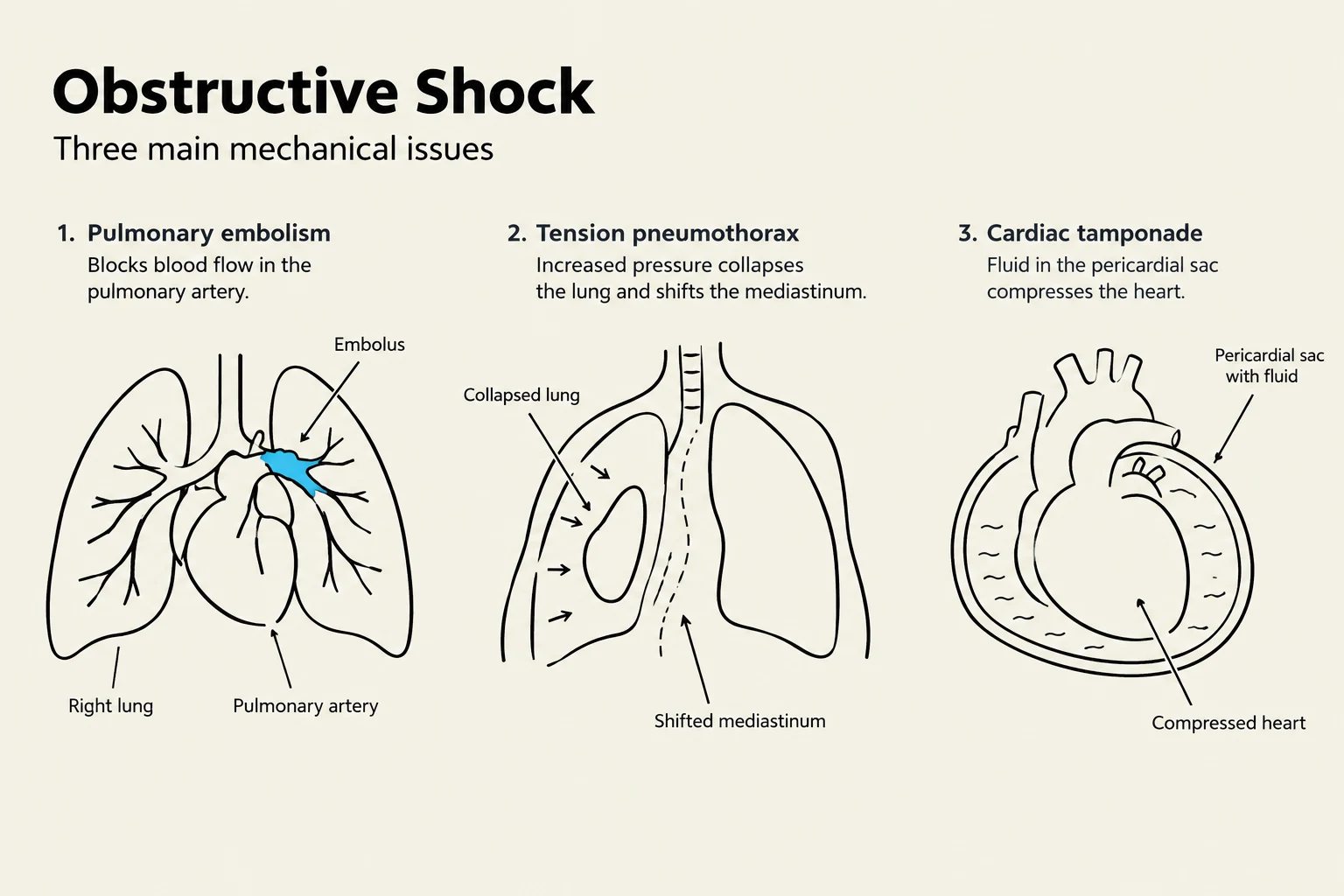
3. Obstructive Shock: "The Blockage"
Obstructive shock is caused by a mechanical obstruction within the circulatory system that prevents blood from flowing through the heart or great vessels.
Common Causes:
Pulmonary Embolism: A blood clot in the lungs (the most common cause).
Cardiac Tamponade: Fluid buildup in the pericardial sac compressing the heart.
Tension Pneumothorax: Air trapped in the chest cavity collapsing the lung and shifting structures.
Dissecting Aortic Aneurysm.
Diagram illustrating three main mechanical causes of obstructive shock: pulmonary embolism, tension pneumothorax, and cardiac tamponade.
4. Distributive Shock: "Blood in the Wrong Place"
In distributive shock, the total blood volume may be normal, but there is a massive loss of vascular tone (vasodilation). This causes blood to "pool" in the periphery rather than returning to the heart. This category includes three distinct subtypes:
Septic Shock
Sepsis is a suspected or proven infection accompanied by a Systemic Inflammatory Response Syndrome (SIRS), characterized by fever, tachycardia, increased white blood cell count, and altered LOC. Septic shock is the most severe form, where hypotension persists despite adequate fluid resuscitation.
The pathophysiology involves a massive release of inflammatory chemicals that cause widespread vasodilation, increased vascular permeability (causing fluid to leak out of vessels), and an inappropriately activated coagulation cascade.
Clinical Signs:
Warm, flushed skin (due to vasodilation)
Fever and increased leukocytes
Elevated serum lactate (a marker of anaerobic metabolism)
Anaphylactic Shock
This is an extreme immune overreaction to an allergen (e.g., food, medication, or insect stings). It is characterized by wheezing, coughing, itching, urticaria (hives), and life-threatening laryngeal edema or bronchospasm.
Neurogenic Shock
Unlike most other forms of shock, neurogenic shock often presents with bradycardia (slow heart rate) and warm, dry skin. This is caused by a loss of sympathetic control over blood vessel tone, often due to spinal cord or brain injury.
Complications and Systemic Failure
If the underlying cause of shock is not corrected promptly, the resulting lack of oxygen leads to devastating systemic complications:
Acute Lung Injury (ALI) & ARDS: Severe respiratory failure and hypoxemia.
Acute Renal Failure: Due to prolonged lack of blood flow to the kidneys.
Disseminated Intravascular Coagulation (DIC): A medical emergency where the coagulation cascade is activated throughout the entire body, causing simultaneous widespread clotting and bleeding.
Multiple Organ Dysfunction Syndrome (MODS): The final stage where multiple organ systems fail due to prolonged ischemia.
This diagram illustrates the progression from initial shock to systemic complications, detailing how inadequate perfusion leads to cellular dysfunction, organ dysfunction, and ultimately multi-organ failure and increased mortality risk.
Clinical Pearl: In anaphylactic shock, the patient should be placed in a supine position. Sitting up can cause blood to pool in the extremities, potentially accelerating circulatory collapse and death.
Keep reading for free.
Create an account to finish this reading and make it part of your study routine.