


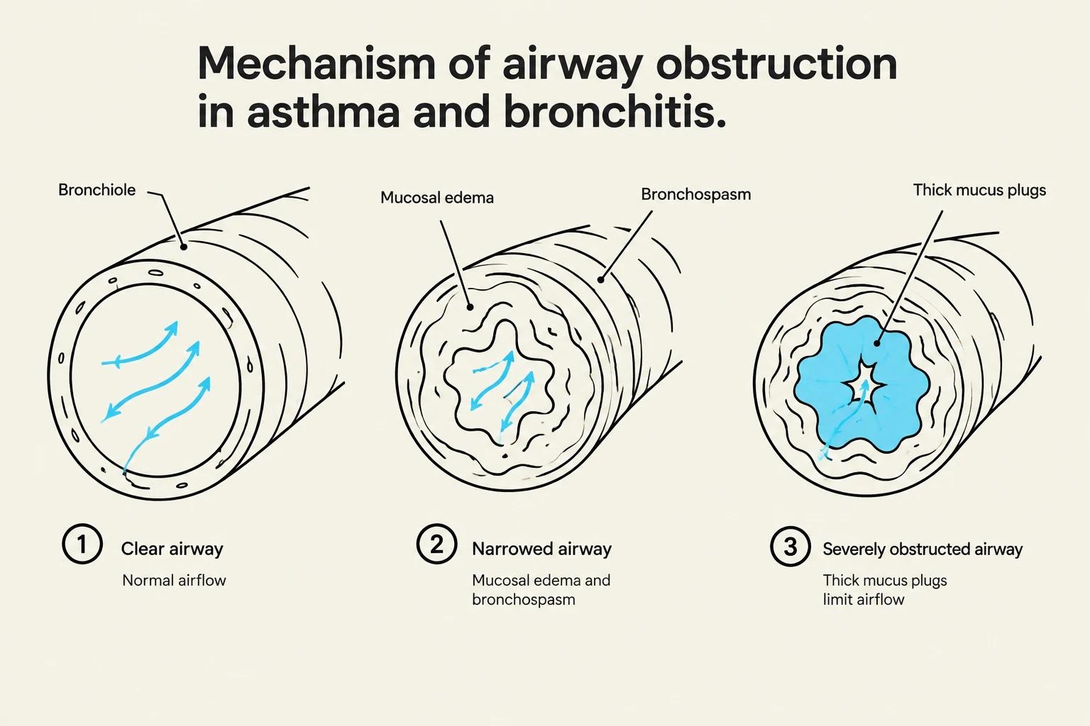
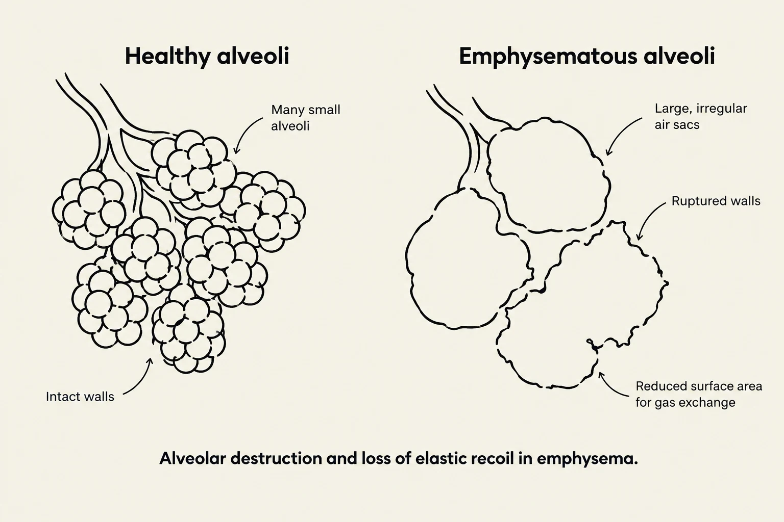
Pulmonary disorders are broadly categorized into two main types based on how they affect lung mechanics and airflow: restrictive and obstructive. While both types can lead to significant respiratory distress, they represent fundamentally different physiological challenges. Restrictive disorders make it difficult to get air into the lungs, whereas obstructive disorders make it difficult to get air out.
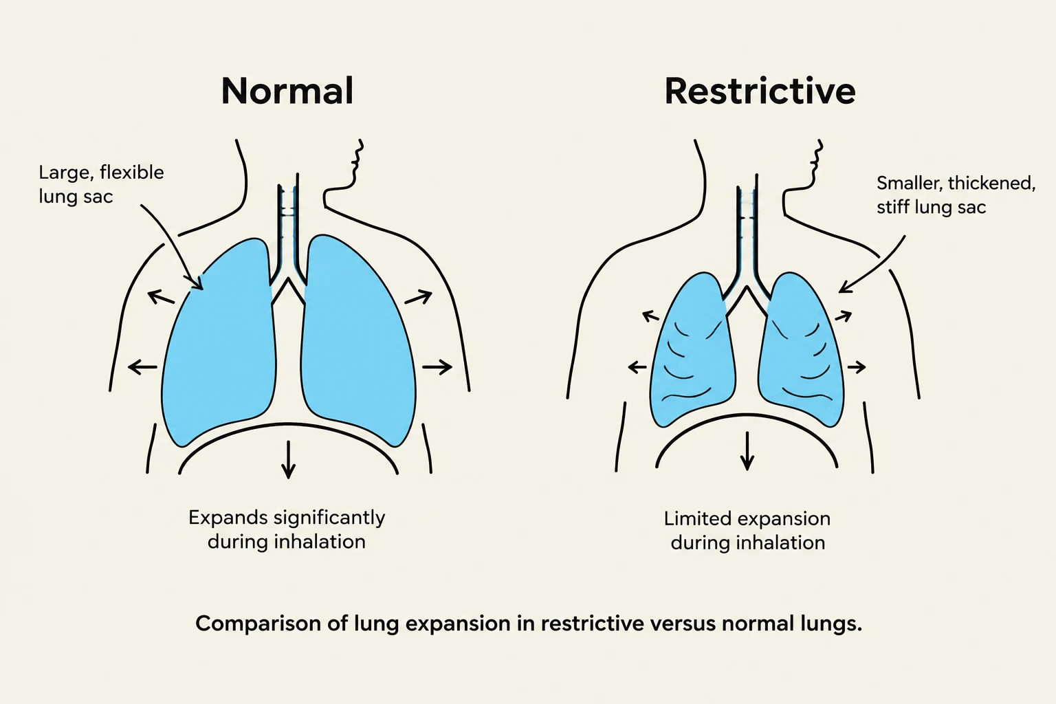
Understanding these distinctions is critical for clinical diagnosis, as they are differentiated through pulmonary function tests, specifically the ratio of forced expiratory volume in one second () to forced vital capacity (FVC).
Restrictive Pulmonary Disorders
Restrictive lung disorders are characterized by a reduction in lung volumes due to the inability of the lungs to fully expand. This is often caused by decreased lung compliance (increased stiffness), a stiff chest wall, or respiratory muscle weakness.
Common Causes and Conditions
- Aspiration: This involves the passage of fluids or solid particles into the lungs. The right lower lobe is the most frequent site of aspiration, and it typically presents with a sudden onset of choking or an intractable cough.
- Atelectasis: This is the collapse of lung tissue. It can be caused by external compression, the gradual absorption of air from hypo-ventilated alveoli, or surfactant impairment (decreased production or inactivation of surfactant).
- Pulmonary Fibrosis: This condition involves the accumulation of excessive fibrous or connective tissue in the lungs. Patients often experience increasing dyspnea (shortness of breath) on exertion.
- Pneumoconiosis: These are lung changes caused by the inhalation of inorganic dust particles, often in a workplace setting. Common examples include diseases caused by silica, asbestos, or coal (such as "Black lung").
- Pulmonary Edema: This is the presence of excess water in the lungs due to disturbances in capillary hydrostatic pressure, oncotic pressure, or permeability. Left-sided heart disease is the most common cause.
- Acute Respiratory Distress Syndrome (ARDS): A severe form of acute lung injury characterized by diffuse alveolocapillary damage, increased capillary permeability, and surfactant inactivation. A hallmark of ARDS is hypoxemia that responds poorly to supplemental oxygen.
Keep reading for free.
Create an account to finish this reading and make it part of your study routine.
Continue reading freeAlready have an account? Log in
Updated