


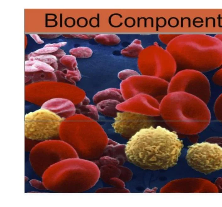
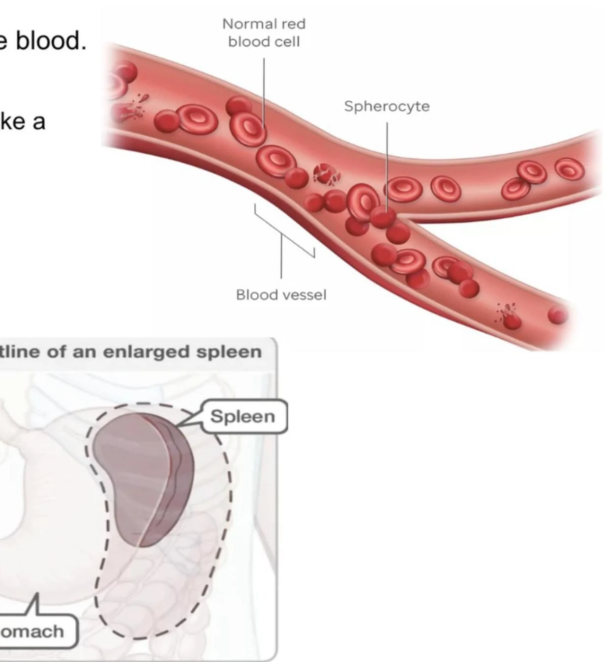
Red blood cells (erythrocytes) are specialized, biconcave discs designed for a single, vital mission: transporting oxygen from the lungs to the body's tissues and carrying carbon dioxide back to be exhaled. To perform this task effectively, these cells must be highly flexible, allowing them to squeeze through even the narrowest capillaries. When the structure or composition of these cells is compromised, the entire circulatory system can suffer. Two significant disorders that illustrate this vulnerability are sickle cell anemia and spherocytosis.
The Normal Erythrocyte
Under normal conditions, erythrocytes are small, flexible, and lack a nucleus or organelles, which maximizes the space available for hemoglobin. Hemoglobin is the protein within the cell responsible for binding oxygen. These cells typically survive for approximately 100 to 120 days in circulation, after which they are removed and recycled by macrophages in the spleen and liver.
Sickle Cell Anemia: A Genetic Mutation
Sickle cell anemia is not a defect of the cell's outer membrane, but rather a defect in the hemoglobin itself. It is caused by a specific genetic mutation in the hemoglobin chain.
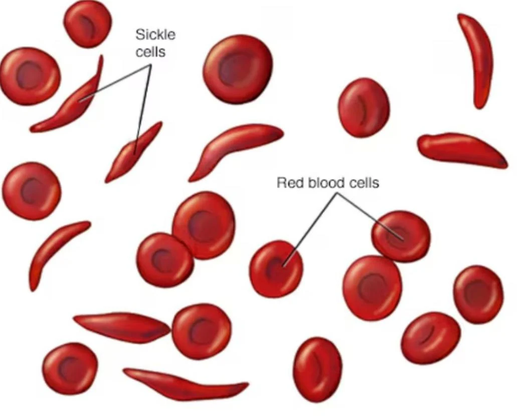
In a healthy individual, the sixth amino acid in this chain is glutamic acid. However, in patients with sickle cell anemia, this is replaced by valine. This single amino acid substitution changes the chemical properties of the hemoglobin molecule, causing the molecules to assemble into long, rigid fibers when oxygen levels are low.
These long fibers distort the normally flexible, biconcave red blood cells into rigid, crescent-shaped sickle cells.

Keep reading for free.
Create an account to finish this reading and make it part of your study routine.
Continue reading freeAlready have an account? Log in
Updated