An in-depth explanation of red blood cells (RBCs), their function in oxygen transport, and the various types of anemia, including hemolytic anemia, iron deficiency anemia, and sickle cell disease. Covers mechanisms, inherited and acquired causes, clinical crises, diagnosis, and laboratory evaluation of RBC indices.
Red blood cells (RBCs), or erythrocytes, are the most numerous formed elements in the blood. Their primary function is to carry oxygen throughout the body, a task facilitated by their unique biconcave disc structure, which provides a large surface area and reversible deformability. When the body's ability to transport oxygen is compromised—either through blood loss, impaired production, or increased destruction—the result is anemia.
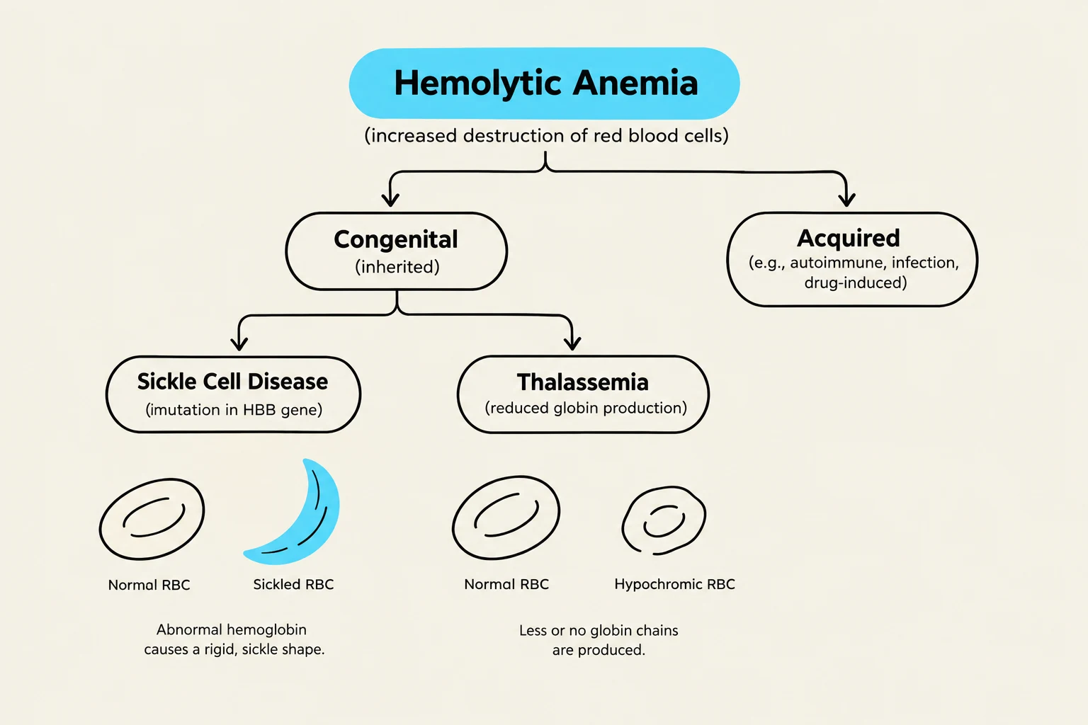
While many forms of anemia exist, such as iron deficiency or vitamin B12 deficiency, a specific and serious category is hemolytic anemia. This condition is characterized by the early destruction of RBCs. These disorders can be categorized by their underlying mechanism: congenital (inherited) or acquired.
Hemolytic Anemia: Mechanisms and Types
Hemolytic anemia occurs when the lifespan of the RBC is significantly shortened. Because the cells are being destroyed faster than the bone marrow can replace them, patients often present with signs of hypoxia (low oxygen), such as fatigue, pallor, and weakness, along with compensatory increases in heart rate and respiratory rate.
Inherited Hemolytic Disorders
Inherited forms of hemolytic anemia are often recessive genetic diseases that affect the structure of the cell or the hemoglobin within it. Key examples include:
Hereditary Spherocytosis: A condition where the RBC membrane is defective, causing the cells to become spherical rather than biconcave, making them more fragile.
Thalassemia: A genetic disease characterized by abnormal hemoglobin production, often resulting in microcytic-hypochromic cells (small, pale cells).
Sickle Cell Disease: A severe form of inherited anemia caused by a mutation in the β chain of the hemoglobin molecule.
Acquired Hemolytic Disorders
Acquired hemolysis is not due to genetics but rather external factors that damage the cells. These include:
Immune-mediated causes: Autoimmune reactions or transfusion reactions.
Mechanical causes: Damage from prosthetic heart valves.
Infections and drugs: Various chemical or biological triggers.
Exam Tip: When evaluating hemolytic anemia, look for clinical signs of jaundice and an increased reticulocyte count. An increased reticulocyte count indicates that the bone marrow is working overtime to replace the rapidly destroyed RBCs.
This diagram illustrates the classification of hemolytic anemia, distinguishing between congenital (inherited) and acquired forms. It further details specific congenital conditions like Sickle Cell Disease and Thalassemia, showing their associated red blood cell morphologies and underlying causes.
Sickle Cell Disease (SCD)
Sickle cell disease is a profound example of an inherited hemolytic disorder. It is caused by the presence of Hemoglobin S (Hb S), an abnormal form of hemoglobin resulting from a genetic mutation.
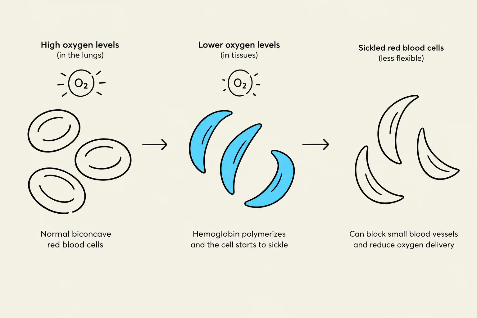
While individuals with "Sickle Cell Trait" (heterozygotes) usually have milder symptoms, those with Sickle Cell Disease (homozygotes) suffer from significant physiological complications. When oxygen levels decrease (hypoxia), the Hb S causes the RBCs to undergo sickling. These sickled cells are rigid and cannot deform easily to pass through small capillaries.
The Clinical Crises of SCD
The sickling process leads to several life-threatening "crises":
Vaso-occlusive Crisis: The rigid, sickled cells block blood vessels, leading to intense pain and tissue infarction (death) in areas like the bones, abdomen, spleen, or brain.
Acute Chest Syndrome: A severe complication presenting with chest pain, fever, cough, and shortness of breath, often caused by pulmonary infarction.
Sequestration: This occurs when blood "pools" in the spleen, leading to sudden spleen enlargement and potentially fatal hypovolemia or shock.
Aplastic Crisis: Often triggered by a viral infection like Parvovirus B19, this causes a sudden cessation of RBC production in the bone marrow.
Hyperhemolytic Crisis: A rare event where the rate of RBC destruction is drastically accelerated, often due to infection or certain drugs.
This diagram illustrates the process of red blood cell sickling due to lower oxygen levels, which can lead to blockages in small blood vessels and reduced oxygen delivery.
Diagnosis and Management
Diagnosis is typically confirmed through Hgb electrophoresis, which identifies the presence of Hemoglobin S. While there is currently no cure, management focuses on preventing sickling episodes by avoiding triggers like cold, dehydration, and stress, as well as using blood transfusions or medications like hydroxyurea to decrease the production of abnormal hemoglobin.
Laboratory Evaluation of RBC Indices
To understand the nature of an anemia, clinicians look at erythrocyte indices. These values help categorize the anemia by cell size (MCV) and hemoglobin concentration (MCHC).
Measures the concentration of Hb (Hypochromic = low)
Reticulocyte Count
Immature RBC count
Indicates the rate of RBC production
For example, in Iron Deficiency Anemia, which is a microcytic-hypochromic anemia, you would expect to see a low MCV and low MCHC. Conversely, in Vitamin B12 or Folate Deficiency, you would see a macrocytic anemia due to impaired DNA synthesis during RBC maturation.
This chart illustrates how Mean Corpuscular Volume (MCV) and Mean Corpuscular Hemoglobin Concentration (MCHC) vary in different types of anemia, including microcytic, normocytic, macrocytic, and hemolytic anemia. It also provides a key feature for each type and visual representations of red blood cells.
Keep reading for free.
Create an account to finish this reading and make it part of your study routine.