


In radiographic imaging, achieving a high-quality diagnostic image requires uniform exposure across the entire image receptor (IR). However, human anatomy is rarely uniform; body parts often possess significant variations in thickness and density. When an X-ray beam passes through a patient, these variations cause uneven X-ray attenuation, where thicker or denser tissues absorb more photons than thinner or less dense areas. Without intervention, this results in poor image uniformity, characterized by overexposed (too dark) areas in thin regions and underexposed (too light) areas in thick regions.
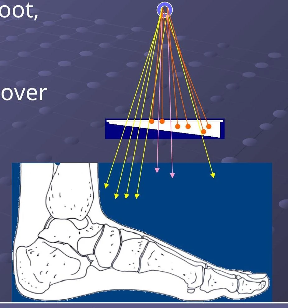
Compensating filters are specialized tools designed to solve this problem. By intentionally attenuating the X-ray beam unevenly, these filters counteract the natural differences in tissue attenuation to provide even radiographic exposure across the image. These filters are typically attached to the face of the collimator, which blocks the field light when the filter is in place.
The Trough Filter
The trough filter is a specific type of compensating filter primarily utilized for AP (Anteroposterior) or PA (Posteroanterior) chest radiography.
The chest presents a unique challenge because the lungs are air-filled and relatively low in density, while the mediastinum (the central compartment containing the heart) and the spine are much denser. To compensate for this, the trough filter is designed with a thicker section of material placed over the lungs and a thinner section positioned over the mediastinum and spine.
Keep reading for free.
Create an account to finish this reading and make it part of your study routine.
Continue reading freeAlready have an account? Log in
Updated