


In emergency medicine, one of the most deceptive scenarios a clinician can face is seeing a rhythmic, organized waveform on a cardiac monitor while the patient remains pulseless. This phenomenon is known as Pulseless Electrical Activity (PEA). While the "wiring" of the heart appears to be functioning correctly, the "pump" itself has failed to generate enough mechanical force to circulate blood through the body.
Defining PEA: The Disconnect Between Electricity and Mechanics
The defining characteristic of PEA is the presence of an organized electrical rhythm on an ECG despite the absence of a palpable pulse. In a healthy heart, electrical signals trigger the myocardium (the heart muscle) to contract, creating a pressure wave we feel as a pulse. In PEA, this connection is broken. The electrical signal may show normal P-waves, QRS complexes, and T-waves, but the myocardium cannot produce sufficient force to move blood.
This is often described as "electromechanical dissociation." The electrical system is working, but the mechanical pump is not.
Clinical Alert: Never rely solely on the monitor. A monitor may look deceptively organized, showing a perfect rhythm, but you must always physically check the patient for a pulse to rule out PEA.
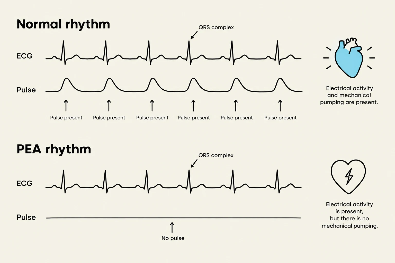
Analyzing the ECG Data
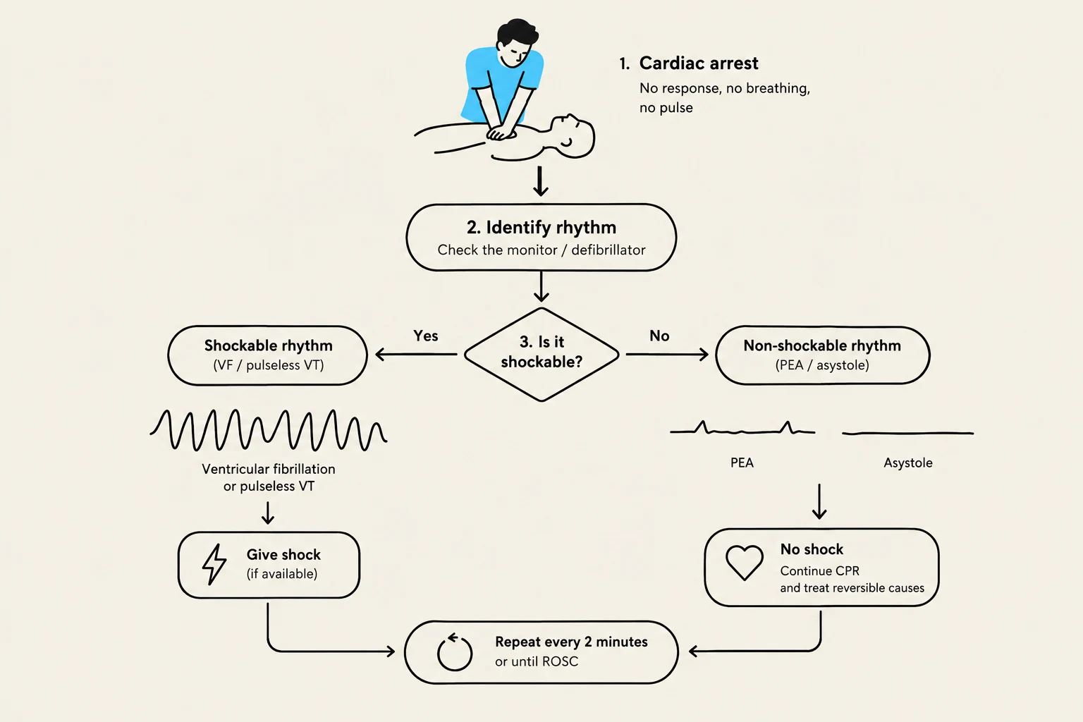
When evaluating a patient in cardiac arrest, the ECG provides vital clues about the type of arrest occurring. PEA is categorized alongside asystole as a "non-shockable" rhythm.

Keep reading for free.
Create an account to finish this reading and make it part of your study routine.
Continue reading freeAlready have an account? Log in
Updated