


Pulmonary edema is a critical medical condition characterized by the accumulation of fluid within the alveoli. When the air sacs of the lungs fill with fluid instead of air, the body's ability to perform efficient gas exchange is severely impaired. While the end result—respiratory distress—looks remarkably similar in many patients, the underlying physiological mechanism is the key to life-saving treatment. To provide effective nursing care, one must distinguish between a "pressure" problem and a "leakage" problem.
The Shared Presentation: Respiratory Distress
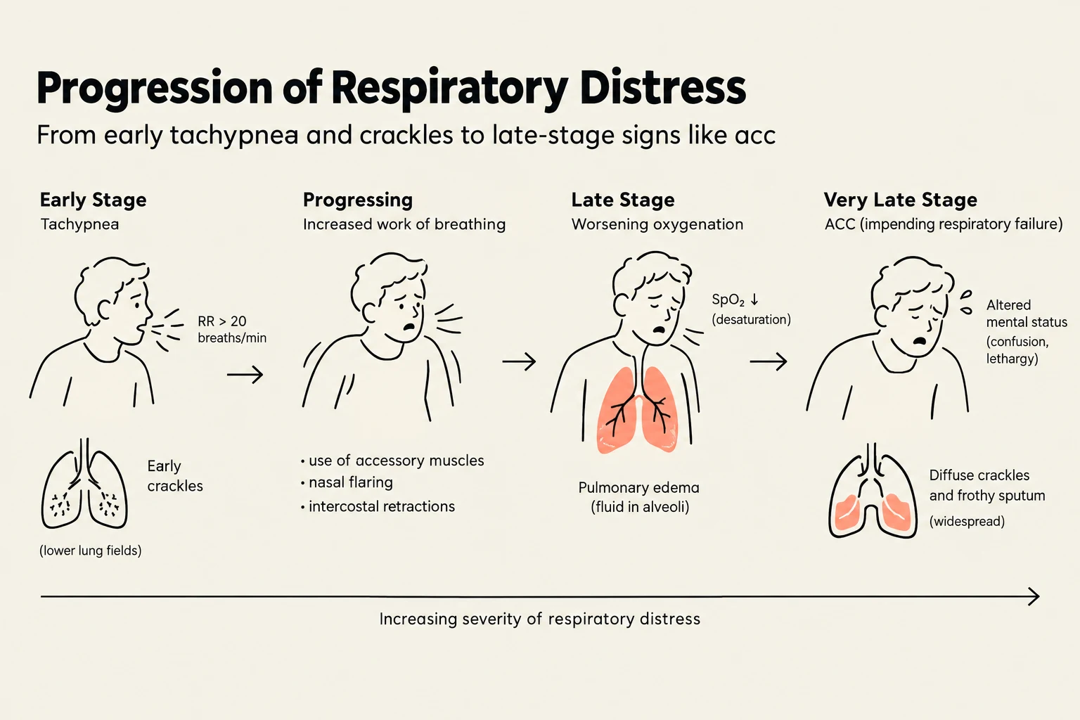
Regardless of whether the cause is the heart or the lungs, a patient in pulmonary edema will present with a common set of alarming symptoms.
Early signs typically include:
- Dyspnea (shortness of breath) and tachypnea (rapid breathing).
- Orthopnea (difficulty breathing while lying flat).
- Crackles or rales heard upon auscultation.
- Anxiety or restlessness due to hypoxia.
As the condition progresses, the patient enters severe respiratory distress. You will observe an increased work of breathing, characterized by:
- Nasal flaring and the use of accessory muscles (e.g., neck and intercostal muscles).
- Retractions (the skin pulling in around the ribs during inhalation).
- Difficulty speaking in full sentences.
🚨 RED FLAG: The "Silent Chest" If a patient who was previously wheezing or struggling suddenly becomes quiet, do not assume they are improving. This may indicate that they are becoming too exhausted to move air at all, signaling imminent respiratory arrest.
Cardiogenic Pulmonary Edema: The "Pressure Cooker"
Cardiogenic pulmonary edema is fundamentally a pump problem. It occurs when the left side of the heart fails to pump blood forward effectively, often due to left-sided heart failure, coronary artery disease, or an acute myocardial infarction (MI).
Keep reading for free.
Create an account to finish this reading and make it part of your study routine.
Continue reading freeAlready have an account? Log in
Updated