Explore the fundamental building blocks of proteins, amino acids, and their role in forming complex 3D structures. Learn about the four levels of protein structure, the importance of folding, and the consequences of misfolding, including prion diseases.
Proteins are the versatile "molecular machines" of the cell. While other macromolecules like lipids and nucleic acids serve as energy stores or information carriers, proteins perform nearly all active cellular tasks. From catalyzing essential biochemical reactions as enzymes to acting as hormones for signaling or antibodies for defense, the functionality of a protein is inseparable from its physical form. The central dogma of protein biology is that a protein's specific function is strictly determined by its unique three-dimensional (3D) structure.
The Building Blocks: Amino Acids
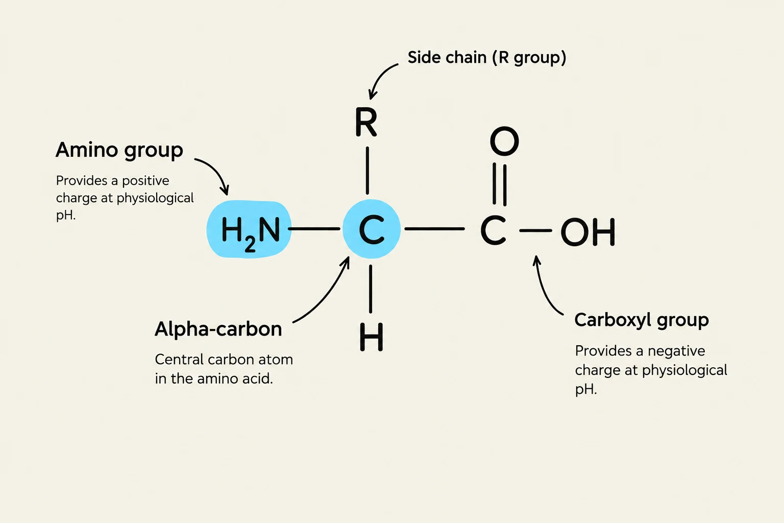
The foundation of every protein is a polymer of amino acids. At physiological pH, each amino acid possesses a consistent core structure: a central α-carbon atom bonded to an amino group (−NH3+), a carboxyl group ($-COO^-$), and a variable R group (side chain).
The R group is the most critical component for a protein's identity. It is this side chain that dictates how an amino acid will interact with its neighbors and its environment. There are 20 standard amino acids, which can be categorized by the chemical nature of their R groups:
Polar Charged: These can form ionic bonds. For example, Aspartate and Glutamate are negatively charged, while Lysine and Arginine are positively charged.
Polar Uncharged: These possess partial charges that allow them to form hydrogen bonds, such as Serine.
Nonpolar: These hydrophobic side chains lack oxygen or nitrogen and tend to cluster away from water, stabilizing the protein's interior via van der Waals forces.
Special Cases:
Glycine is exceptionally small and flexible.
Proline is rigid and often introduces bends in the chain.
Cysteine contains a sulfhydryl ($-SH$) group capable of forming covalent disulfide bonds ($-S-S-$) that provide significant structural stability.
Exam Tip: Remember that the charge of certain amino acids, like Histidine, can change depending on the local chemical environment. Additionally, notice how the positive charge of Histidine-rich proteins (like histones) allows them to bind to the negatively charged phosphate backbone of DNA.
Diagram illustrating the general structure of an amino acid, highlighting the amino group, alpha-carbon, carboxyl group, and side chain (R group).
Linking the Chain: Peptide Bonds and Modifications
Amino acids are joined together through peptide bonds, which form between the carboxyl group of one amino acid and the amino group of the next. This creates a continuous polypeptide chain. While the average polypeptide contains about 300 amino acids, the complexity of a protein can be further increased through protein modifications. After synthesis, proteins may have functional groups added, such as carbohydrates (forming glycoproteins), metals (forming metalloproteins), or flavins (forming flavoproteins).
The Four Levels of Protein Structure
To transform from a simple string of amino acids into a functional machine, a protein must fold into a specific hierarchy of organization.
1. Primary Structure
The primary structure is the linear sequence of amino acids in the polypeptide chain. This sequence is the blueprint for all subsequent folding; even a single mutation in this sequence can disrupt the entire protein's function.
2. Secondary Structure
Secondary structure refers to local, repetitive conformations of the polypeptide backbone. These shapes are stabilized by noncovalent interactions, specifically hydrogen bonds between the atoms of the backbone. The two most common motifs are:
α-helices: A coiled, spring-like structure.
β-sheets: Extended strands that lie side-by-side.
3. Tertiary Structure
The tertiary structure is the full 3D conformation of a single polypeptide chain. This level of folding is driven by various interactions between the R groups, including hydrophobic interactions, ionic bonds, hydrogen bonds, and covalent disulfide bridges.
Proteins are often categorized by their tertiary shape:
Fibrous proteins: Elongated and used for structural roles (e.g., keratin, elastin).
Globular proteins: Compact and functional, often found within the cell (e.g., myoglobin).
Within these structures, you may find domains—compact regions that can fold and function independently.
4. Quaternary Structure
Some proteins consist of multiple polypeptide chains, known as subunits, working together as a single functional unit.
Homodimers: Two identical subunits.
Heterodimers: Two different subunits.
A classic example is hemoglobin, which utilizes four subunits to function. These subunits may be held together by noncovalent bonds or covalent disulfide bonds.
Folding, Chaperones, and Denaturation
The process of a protein finding its lowest-energy, functional state is often driven by hydrophobic collapse, where nonpolar side chains cluster in the center to avoid water. However, the cellular environment is crowded, and folding can be difficult.
To prevent errors, cells use molecular chaperones:
Hsp70 family: These bind to newly emerging polypeptides on ribosomes to shield them from inappropriate interactions.
Chaperonins (e.g., TRiC): These provide an isolated "folding chamber" where complex proteins can fold without interference.
If the stabilizing interactions (like hydrogen bonds or hydrophobic contacts) are disrupted by heat, radiation, chemicals, or detergents, the protein undergoes denaturation. A denatured protein loses its 3D shape and, consequently, its ability to function.
When Folding Goes Wrong: Misfolding and Disease
While most proteins fold correctly, misfolding can have deadly consequences. Misfolded proteins can expose hydrophobic surfaces that cause them to stick together, forming stable, insoluble aggregates called amyloid fibrils.
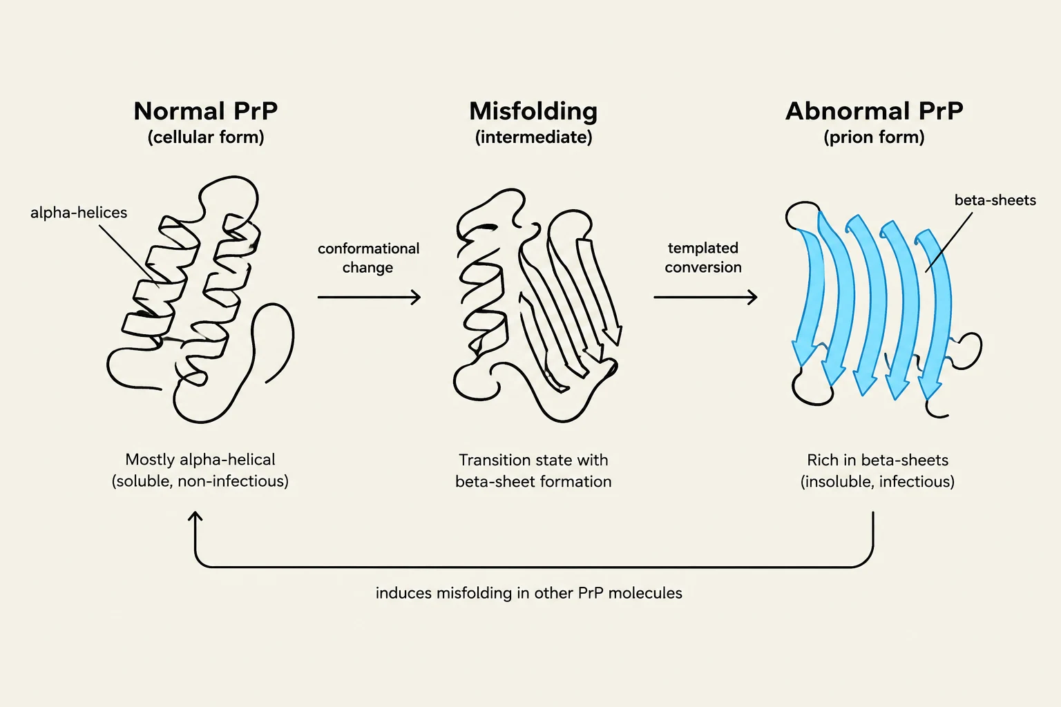
A terrifying example of this is Prion diseases, such as Creutzfeldt–Jakob disease (CJD). In these cases, a normal protein ($PrP^C$) undergoes a conformational change into an abnormal, misfolded state (PrPSc). This abnormal form can act as a template, inducing other normal proteins to misfold, leading to progressive neurodegeneration.
This diagram illustrates the process of normal prion protein (PrP) misfolding into an abnormal, infectious form. The normal cellular form is mostly alpha-helical and soluble, while the abnormal prion form is rich in beta-sheets, insoluble, and infectious. The misfolding process involves a conformational change and can be templated, leading to the induction of misfolding in other PrP molecules.
Summary Note: Protein function is a delicate balance of weak noncovalent bonds and strong covalent bonds. When this balance is tipped by mutation or environmental stress, the results can range from simple loss of function to catastrophic disease.
Keep reading for free.
Create an account to finish this reading and make it part of your study routine.