Understanding Potassium Imbalances: Hyperkalemia and Hypokalemia
This content explains the critical role of potassium in the body, detailing the causes, clinical manifestations, and ECG changes associated with both hyperkalemia (high potassium) and hypokalemia (low potassium). It also covers the relationship between potassium levels and acid-base balance, and the role of the kidneys
Electrolytes are charged particles—either cations (positive) or anions (negative)—that are essential for maintaining fluid balance, generating electrical impulses, and ensuring proper cellular function. Among these, potassium (K+) is the primary intracellular cation, meaning it is concentrated within the cells (Intracellular Fluid, or ICF). Because potassium is critical for nerve impulse conduction, protein synthesis, and, most importantly, the electrical activity of the heart, even minor deviations from the normal range can lead to life-threatening consequences.
The normal serum potassium range for an adult is typically 3.5 to 5.3 mEq/L. Values falling below 2.5 mEq/L or rising above 6.2 mEq/L are considered critical and require immediate medical intervention.
The Role of the Kidney and Acid-Base Balance
The kidneys serve as the most efficient regulator of potassium, managing its excretion to maintain homeostasis. This regulation is heavily influenced by hormones like aldosterone, which promotes potassium excretion in exchange for sodium reabsorption.
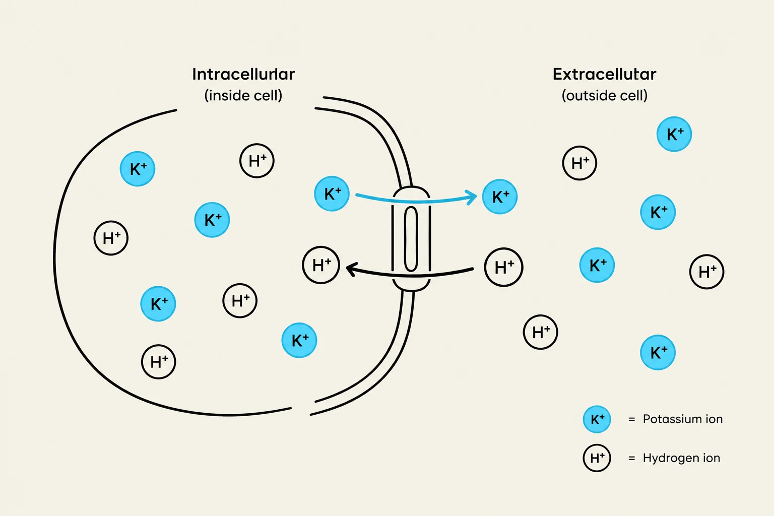
A crucial concept for clinicians is the relationship between potassium and acid-base balance. Potassium levels often shift in response to changes in pH through an exchange mechanism between the intracellular fluid (ICF) and the extracellular fluid (ECF).
Acidosis: When the body is in a state of acidosis (low pH), there is an accumulation of hydrogen ions (H+) in the cells. To maintain electrical balance, H+ moves into the cells, and K+ moves out into the ECF, resulting in hyperkalemia.
Alkalosis: Conversely, in alkalosis (high pH), there is a deficit of H+ in the cells. H+ moves out of the cells to compensate, causing K+ to move into the cells, resulting in hypokalemia.
Diagram illustrating the movement of potassium (K+) and hydrogen (H+) ions across a cell membrane, differentiating between intracellular and extracellular environments.
Hyperkalemia: High Potassium (K+>5.3 mEq/L)
Hyperkalemia occurs when there is too much potassium in the extracellular fluid. This is most commonly seen in patients with renal failure, as the kidneys can no longer effectively excrete the excess.
Causes of Hyperkalemia
A helpful mnemonic for remembering the causes of increased serum potassium is "MACHINES":
Medications (e.g., ACE inhibitors, NSAIDs)
Acidosis (Metabolic or respiratory)
Cellular destruction (Burns, traumatic injury)
Hypoaldosteronism, Hemolysis
Intake (Excessive)
Nephrons (Renal failure)
Excretion (Impaired)
Clinical Manifestations
The symptoms of hyperkalemia depend on the severity of the elevation:
Mild attacks: Increased neuromuscular irritability, restlessness, intestinal cramping, and diarrhea.
Severe attacks: Decreased resting membrane potential leading to muscle weakness, loss of muscle tone, and flaccid paralysis.
Electrocardiograph (ECG) Changes
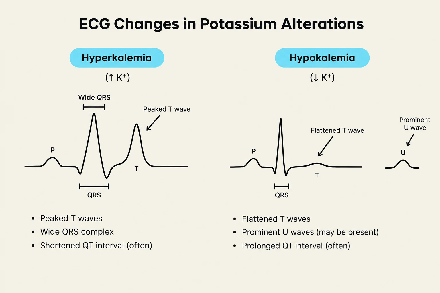
Hyperkalemia is a cardiac emergency because it disrupts the electrical conduction of the heart. Common ECG changes include:
Tall, peaked T waves
Prolonged PR intervals
Flat or absent P waves
Widened QRS complex
Bradycardia and heart blocks
Critical Risk: Severe hyperkalemia can lead to ventricular fibrillation or cardiac standstill.
Exam Tip: If a patient presents with peaked T waves and a history of renal failure, suspect hyperkalemia immediately. Treatment may include calcium gluconate to stabilize the heart, or insulin and glucose to shift potassium back into the cells.
Hypokalemia: Low Potassium (K+<3.5 mEq/L)
Hypokalemia is characterized by a decrease in extracellular potassium levels. This is often caused by a loss of total body potassium through gastrointestinal or renal routes.
Common Causes
Gastrointestinal loss: Vomiting, diarrhea, or excessive use of laxatives.
Renal loss: Use of diuretics (specifically those that increase urinary output) or glucocorticoids.
Cellular shifts: Insulin administration promotes the uptake of potassium into the cells, which can lower ECF levels.
Clinical Manifestations
When managing hypokalemia, clinicians often use the phrase "Think SLOW" to remember the systemic impact:
Skeletal muscle weakness and cramps
Lethargy and fatigue
Oliguria (low urine output)
Weakness of smooth muscles, leading to decreased intestinal motility (constipation, nausea, or paralytic ileus).
Electrocardiograph (ECG) Changes
Hypokalemia slows down electrical conduction, leading to distinct ECG patterns:
Flattened or shallow T waves
Prominent U waves
ST segment depression
Premature ventricular contractions (PVCs) and ventricular tachycardia.
This diagram illustrates the characteristic electrocardiogram (ECG) changes associated with hyperkalemia (high potassium levels) and hypokalemia (low potassium levels), highlighting differences in T waves, QRS complexes, and the presence of U waves.
Summary of Manifestations
The following table summarizes the key differences in how potassium alterations impact various organ systems:
Organ System
Hypokalemia (K+↓)
Hyperkalemia (K+↑)
Cardiovascular
Dysrhythmias, flattened T waves, U waves, ST depression, cardiac arrest
Dysrhythmias, peaked T waves, prolonged PR, wide QRS, cardiac arrest
Clinical Note: Long-term potassium deficits (lasting more than one month) can lead to permanent kidney damage, including interstitial fibrosis and tubular atrophy.
Keep reading for free.
Create an account to finish this reading and make it part of your study routine.