Understanding Oxygenation and Ventilation Disorders: Asthma and COPD
This document provides a comprehensive overview of oxygenation and ventilation disorders, focusing on the pathophysiology, clinical manifestations, diagnostic tools, and management strategies for Asthma and Chronic Obstructive Pulmonary Disease (COPD). It also includes guidance on nursing management and patient educati
Effective breathing is a complex physiological process requiring the seamless coordination of anatomical structures, neurological signals, and gas exchange mechanisms. When this process is disrupted, patients experience oxygenation and ventilation disorders. These disorders can stem from various origins, including anatomical abnormalities like a deviated septum, infections and inflammation of the airway linings, physical obstructions such as tumors or foreign bodies, or neurological dysfunctions that impair the muscles responsible for breathing.
Among the most prevalent ventilation disorders are Asthma and Chronic Obstructive Pulmonary Disease (COPD). While they share several clinical features, their underlying pathophysiology and long-term management strategies differ significantly.
Understanding Oxygenation-Ventilation Disorders
To understand respiratory disease, one must first categorize the types of disruptions that can occur in the respiratory system:
Anatomical Issues: Physical abnormalities in the structure of the respiratory tract, such as polyps or a deviated septum, which restrict airflow.
Infection and Inflammation: Immune responses to bacterial, viral, or fungal pathogens that cause swelling and excess mucus.
Foreign Bodies or Tumors: Physical obstructions caused by inhaled objects or abnormal tissue growths.
Neurological or Physical Factors: Dysfunction in the nerves controlling airway muscles or external pressure affecting the mechanics of breathing.
Asthma: Pathophysiology and Clinical Manifestations
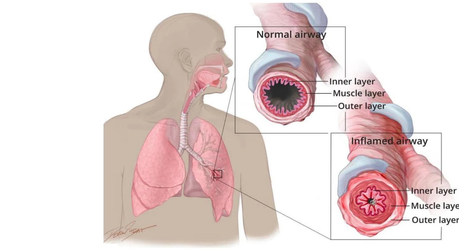
Asthma is defined as a chronic inflammatory condition of the bronchial tubes. It is characterized by increased sensitivity of the airways, which leads to breathing difficulties when triggered by genetics, allergies, or environmental factors like smoke and dust.
Diagram illustrating the difference between a normal airway and an inflamed airway, showing the layers of the airway wall.
The pathophysiology of an asthma attack involves three primary mechanisms:
Airway Inflammation: The lining of the airways becomes swollen and irritated.
Airway Constriction: The smooth muscle mass surrounding the airways tightens, narrowing the passage.
Increased Mucus Production: Excessive secretion further obstructs the already narrowed lumen.
Clinical Presentation
Asthma typically manifests in childhood or early adulthood and is often acute and episodic, meaning the airflow obstruction is frequently reversible with treatment. Common symptoms include:
Shortness of breath (dyspnea) and orthopnea (difficulty breathing while lying flat).
Chest tightness and persistent coughing.
Expiratory wheezing (though severe cases may present with inspiratory wheezing or diminished breath sounds).
Systemic signs of distress: Tachycardia, diaphoresis (sweating), anxiety, panic, and central cyanosis.
Exam Tip: If a patient with asthma presents with a sudden absence of wheezing accompanied by severe intercostal retractions, this may indicate a life-threatening decline in airflow rather than improvement.
Diagnostic Tools
To confirm an asthma diagnosis, clinicians utilize several tools, including Chest X-rays (CXR), Arterial Blood Gas (ABG) analysis, Serum IgE levels, and allergy skin testing.
Status Asthmaticus: A Medical Emergency
Status asthmaticus is an acute, extreme exacerbation of asthma. It is a medical emergency characterized by hypoxemia (low blood oxygen) and hypercarbia (high blood carbon dioxide) that fails to respond to standard bronchodilators or corticosteroids.
Management of status asthmaticus requires aggressive intervention:
Oxygen Therapy: High-flow oxygen to maintain SpO2 levels between $90%-92%$ or higher.
Pharmacotherapy:
Beta-2 Agonists: Continuous nebulized albuterol or levalbuterol to relax smooth muscles.
Corticosteroids: Early administration of intravenous methylprednisolone or dexamethasone to combat inflammation.
Anticholinergics: Nebulized ipratropium bromide to provide additional airway relaxation.
Mechanical Ventilation: If oxygen saturation continues to decline despite these measures, the patient may require mechanical ventilation.
Chronic Obstructive Pulmonary Disease (COPD)
While asthma involves smooth muscle tightness with relatively intact air sacs, COPD involves permanent, structural lung damage. COPD is an umbrella term that primarily includes Chronic Bronchitis and Emphysema.
Diagram illustrating the difference between healthy lungs and lungs affected by COPD, showing narrowed bronchioles and alveolar membrane breakdown in emphysema.
Emphysema: The "Pink Puffer"
Emphysema is characterized by the destruction of the alveoli (air sacs), leading to enlarged, inefficient air spaces. There are two primary types:
Centriacinar Emphysema: Often caused by heavy smoking; it affects the inner or middle part of the air unit while the outer alveoli remain normal.
Panacinar Emphysema: Often linked to alpha-1 antitrypsin (AAT) deficiency; it affects the entire air unit from front to back, enlarging every single air sac.
A medical illustration of enlarged air sacs characteristic of panacinar emphysema. This illustrates pulmonary pathology.
Patients with emphysema are often referred to as "pink puffers" due to their tendency to maintain relatively normal oxygen levels through increased work of breathing, despite the structural damage.
Chronic Bronchitis: The "Blue Bloater"
Chronic bronchitis involves long-term inflammation of the bronchi, leading to excessive mucus production. These patients are often called "blue bloaters" because they frequently present with:
Cyanosis (bluish skin discoloration).
Cough with sputum production.
Edema and Distended Abdomen: Often due to right-sided heart failure (Cor Pulmonale) resulting from pulmonary hypertension.
Comparative Summary: Asthma vs. COPD
Both conditions share common symptoms such as coughing, wheezing, dyspnea, chest tightness, and difficulty expelling air. They can both be exacerbated by smoke, dust, or cold air and are commonly managed with inhalers.
Feature
Asthma
COPD
Primary Issue
Smooth muscle constriction & inflammation
Structural lung damage (alveoli/bronchi)
Onset
Childhood/Early adulthood; episodic
Typically older adults; progressive
Reversibility
Often reversible with treatment
Generally irreversible
Key Triggers
Allergies, genetics, environmental
Smoke, tobacco, air pollution
Nursing Management and Patient Education
Nursing care for both disorders focuses on maintaining airway patency, monitoring respiratory status, and providing comfort.
For Asthma Patients
Assessment: Monitor for signs of respiratory distress, including use of accessory muscles or altered mental status.
Inhaler Technique: Ensure the patient uses their Metered Dose Inhaler (MDI) correctly. The use of a spacer is highly recommended to improve medication delivery.
Education: Teach the distinction between quick-relief (rescue) medications and long-term control (anti-inflammatory) medications.
For COPD Patients
Positioning: Use High-Fowler's or the Tripod position to facilitate breathing.
Breathing Techniques: Teach pursed-lip breathing, diaphragmatic breathing, and the huff coughing technique to help expel air.
Nutrition: Encourage high-calorie, high-protein meals and high fluid intake (2-3 liters/day) to support energy needs and thin secretions, if not contraindicated.
Energy Conservation: Advise patients to space out activities with scheduled rest periods.
MDI Administration Guide
Proper administration is critical for effective therapy:
Prepare: Shake the puffer for 5-10 seconds and remove the cap.
Prime: If the inhaler is new, spray it into the air first.
Exhale: Breathe out fully away from the device to empty the lungs.
Inhale: Seal lips around the mouthpiece, press the canister once, and breathe in slowly and deeply (not quickly).
Hold: Remove the device and hold the breath for 10 seconds.
Rinse: If using corticosteroids, rinse the mouth with water and spit it out to prevent oral thrush.
Clinical Pearl: Always remind patients that if they are using a corticosteroid inhaler, rinsing their mouth is not optional—it is a vital step to prevent fungal infections (thrush).
Keep reading for free.
Create an account to finish this reading and make it part of your study routine.