Understanding Non-Gynecologic Pelvic Masses and the Postmenopausal Pelvis
This guide explains how to differentiate non-gynecologic pelvic masses from gynecologic ones using sonography. It covers mimics from the GI and urinary tracts, medical devices, and postoperative changes. It also details sonographic findings in the postmenopausal pelvis, including ovarian changes, endometrial thickness
In clinical sonography, distinguishing between gynecologic and non-gynecologic pelvic masses is a fundamental skill. While many pelvic masses originate from the reproductive organs, several other anatomical systems—including the gastrointestinal tract, the urinary tract, and even medical devices—can mimic gynecologic pathology. Furthermore, the physiological shift that occurs during menopause significantly alters the sonographic appearance of the uterus and ovaries, requiring specific diagnostic thresholds to differentiate normal aging from malignancy.
Non-Gynecologic Pelvic Masses
A "mass" in the pelvis is not always a tumor or a cyst of the reproductive system. Sonographers must maintain a high index of suspicion for several non-gynecologic etiologies:
Gastrointestinal and Urinary Tract Mimics
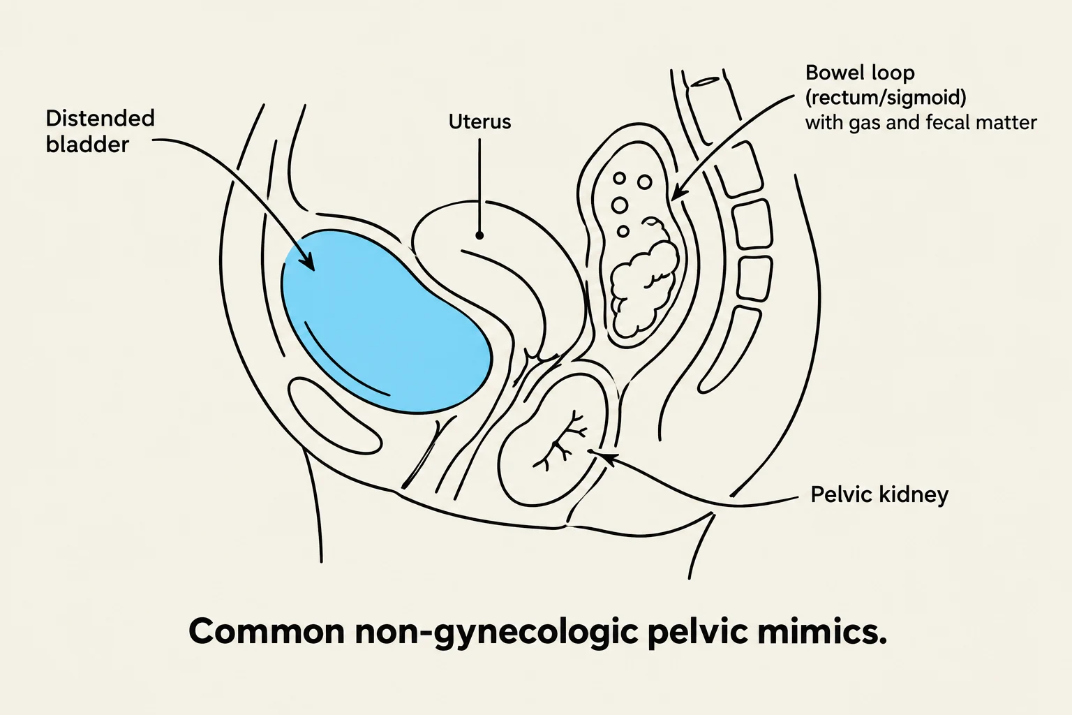
Diagram illustrating common non-gynecologic pelvic mimics, including a distended bladder, uterus, bowel loop with gas and fecal matter, and a pelvic kidney.
The gastrointestinal (GI) tract is a frequent source of "pseudo-masses." An enlarged colon filled with fecal material can simulate a complex mass in the cul-de-sac, while a fluid-filled rectum can mimic a cystic adnexal mass. Additionally, inflammatory conditions like diverticulitis (left-sided) or appendicitis and Crohn’s disease (right-sided) can present as adnexal masses. Carcinomas of the rectosigmoid, cecum, or ileum may also mimic adnexal masses, often appearing with a "target sign" due to central air within the lumen.
The urinary tract also presents challenges. A pelvic kidney can be mistaken for a palpable mass, but it is easily identified by the absence of a kidney in its normal anatomical position. A distended bladder may be mistaken for a cystic ovarian mass, and bladder diverticula or dilated distal ureters can simulate adnexal cysts.
Medical Devices and Postoperative Changes
Comparison of Copper IUCD and Plastic polymer IUCD on ultrasound, illustrating their echogenicity and shadowing characteristics.
Intrauterine Contraceptive Devices (IUCD): These are placed in the uterine cavity for birth control. Copper devices (like the Paraguard) are highly reflective and cause posterior shadowing, while plastic polymer devices (like Mirena) are mildly echogenic.
Vaginal Pessaries: Used to manage pelvic organ prolapse, these devices can create a large shadow in the vaginal and cervical area on both transabdominal and transvaginal ultrasound.
Postoperative Masses: Following pelvic surgery, sonographers may encounter abscesses (ovoid with thick irregular walls), hematomas (varying echogenicity based on age), lymphoceles, urinomas, or seromas.
Ovarian Vein Thrombophlebitis: An uncommon but life-threatening condition, often occurring postpartum or after surgery. It typically involves the right ovarian vein (90% of cases) and appears as a tubular, avascular structure on Doppler.
The Postmenopausal Pelvis
Menopause is defined by the cessation of follicle production in the ovaries because they can no longer respond to Follicle Stimulating Hormone (FSH). This physiological shift leads to a significant decline in the production of estrogen and progesterone, which in turn causes a decrease in the size of both the uterus and the ovaries.
Ovarian Changes
Because they lack functioning follicles, postmenopausal ovaries decrease in size. As a rule of thumb, the ovaries should not exceed 2 cm in diameter or 8 cm3 in volume. Due to natural atrophy, these organs may become difficult to differentiate from the surrounding pelvic structures during sonographic evaluation.
Endometrial Thickness and Postmenopausal Bleeding
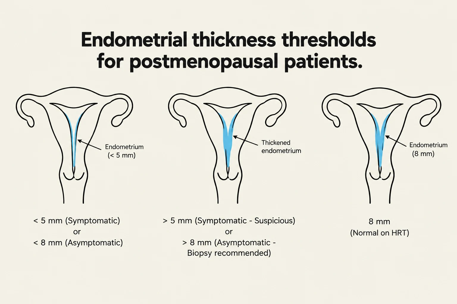
Diagrams illustrating endometrial thickness thresholds for postmenopausal patients, indicating normal, thickened, and suspicious endometrium based on measurements and symptoms.
Sonographic evaluation is critical for patients experiencing postmenopausal bleeding, defined as any vaginal bleeding that is not cyclic (such as that caused by sequential Hormone Replacement Therapy).
The interpretation of endometrial thickness depends heavily on whether the patient is symptomatic (experiencing Abnormal Uterine Bleeding, or AUB) or asymptomatic:
Patient Status
Endometrial Thickness Threshold
Clinical Recommendation
Asymptomatic
<8 mm
Normal
Asymptomatic
>8 mm
Biopsy recommended
Symptomatic (AUB)
<5 mm (homogenous/regular)
Cancer unlikely; normal
Symptomatic (AUB)
>5 mm
Suspicious; recommend SIS or biopsy
Exam Tip: A thin endometrium (typically <5 mm in symptomatic patients) is a strong indicator that excludes endometrial cancer.
Impact of Medications: HRT and Tamoxifen
Medication use can significantly alter the "normal" sonographic appearance of the pelvis, making standard measurements difficult to interpret.
Hormone Replacement Therapy (HRT)
Approximately 30% of women under age 65 use HRT to protect against fractures and improve well-being. Because HRT often involves combined estrogen and progesterone, it can cause cyclic variations in the endometrium and increase uterine size. Consequently, the uterus of a patient on HRT may look similar to that of a menstruating woman. In these specific patients, an endometrial thickness of 8 mm is considered normal.
Tamoxifen
Tamoxifen is an estrogen analogue used in the treatment of breast cancer. While it blocks the effects of estrogen in breast tissue to inhibit cancer growth, it can have different effects on the uterus. Tamoxifen use is associated with:
Endometrial thickening
Polyps and endometrial hyperplasia
Cystic changes
Increased risk of malignancy
Summary Table: Clinical Decision Making
Finding
Context
Action/Interpretation
Thick Endometrium
Asymptomatic
If >8 mm, biopsy
Thick Endometrium
Symptomatic (AUB)
If >5 mm, SIS or biopsy
Thick Endometrium
On HRT
8 mm is considered normal
Large Ovaries
Postmenopausal
Should be <2 cm or 8 cm3
Keep reading for free.
Create an account to finish this reading and make it part of your study routine.