Understanding Neuromuscular Disorders and Muscle Movement Alterations
This content explains the human motor system, differentiating between alterations in muscle tone (hypotonia, hypertonia, spasticity, rigidity) and muscle movement (hyperkinesia, hypokinesia). It details Myasthenia Gravis, a neuromuscular junction disorder, including its pathophysiology, clinical presentation, and medic
The human motor system is a sophisticated loop of sensory input, central integration, and motor output. When this loop is interrupted—whether by damage to the central nervous system (CNS), the peripheral nervous system (PNS), or the communication point between nerves and muscles—the result is a visible alteration in movement. Understanding these alterations requires a clear distinction between changes in muscle tone and changes in the rhythm or quantity of movement.
Understanding Muscle Tone Alterations
Muscle tone refers to the state of tension in a muscle at rest. Pathological changes in tone are generally categorized into two directions: decreased tone (hypotonia) and increased tone (hypertonia).
Hypotonia and Flaccidity
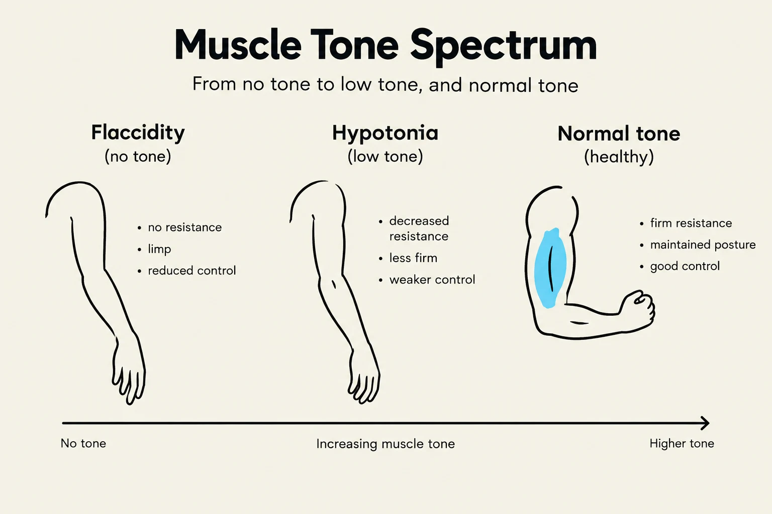
When there is a lack of stimulation due to decreased nerve impulses, such as in a spinal cord injury or a Cerebrovascular Accident (CVA), the muscle exhibits hypotonia. If the nerve impulses are lost entirely, the condition progresses to flaccidity, characterized by a complete lack of muscle tone.
Hypertonia: Increased Muscle Tension
Hypertonia occurs when muscle tone is abnormally increased. This can manifest in several distinct ways depending on the underlying neurological cause:
Spasticity: This is caused by the hyperexcitability of stretch reflexes, often resulting in hyperactive reflexes and clonus.
Paratonia: A specific type of resistance where the force required to move a limb is proportional to the force applied by the examiner.
Dystonia: Characterized by sustained, involuntary twisting movements caused by slow, uninhibited muscle contractions.
Rigidity: This involves tonic reflex activity during passive movement, often described in conditions like Parkinson's disease as "cogwheel" or "lead-pipe" rigidity.
This diagram illustrates the spectrum of muscle tone, ranging from flaccidity (no tone) to hypotonia (low tone) and normal tone (healthy).
Exam Tip: When distinguishing between spasticity and rigidity, remember that spasticity is velocity-dependent (related to the speed of the stretch reflex) and often associated with Upper Motor Neuron (UMN) lesions, whereas rigidity is often constant throughout the range of motion.
Muscle Movement Alterations
While tone refers to tension, movement alterations refer to the actual execution of motor tasks. These are classified by whether there is too much movement (hyperkinesia) or too little movement (hypokinesia).
Hyperkinesia: Excessive Movement
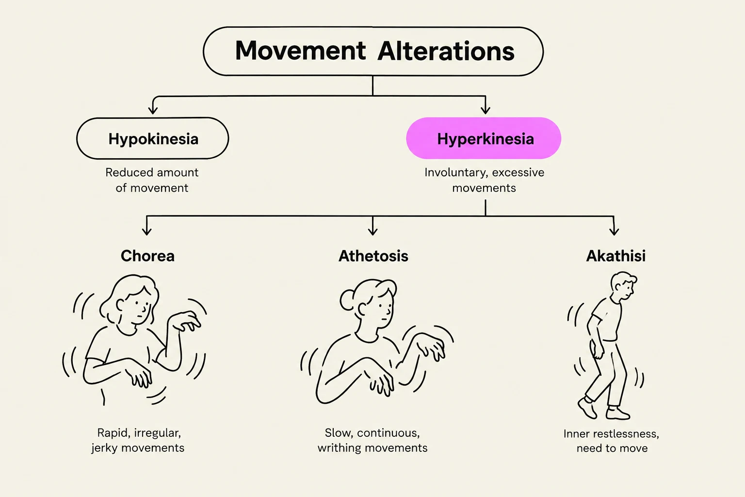
Hyperkinesia involves involuntary, excessive movements. These can be categorized by their pattern:
Chorea: Dysrhythmic, writhing, or "snakelike" muscle contractions.
Athetosis: Slower, more rhythmic, and continuous writhing movements.
Akathisia: A state of hyperactivity driven by a compulsive, distressing desire to move.
Dyskinesia: Abnormal, repetitive, and often stereotypic involuntary movements.
Hypokinesia: Decreased Movement
Hypokinesia involves a reduction in the amount or speed of movement. This includes:
Akinesia: The complete absence of movement.
Bradykinesia: A significant slowing of movement, a hallmark symptom of Parkinson's disease.
This diagram illustrates different types of movement alterations, including Hypokinesia (reduced movement) and Hyperkinesia (excessive movement), with specific examples like Chorea, Athetosis, and Akathisia, each described by their characteristic movements.
Myasthenia Gravis: A Neuromuscular Junction Disorder
While many movement disorders stem from the brain (like Parkinson's) or the spinal cord (like ALS), Myasthenia Gravis (MG) is a specific neuromuscular disease that affects the myoneural junction—the synapse where a motor neuron communicates with a muscle fiber.
Pathophysiology
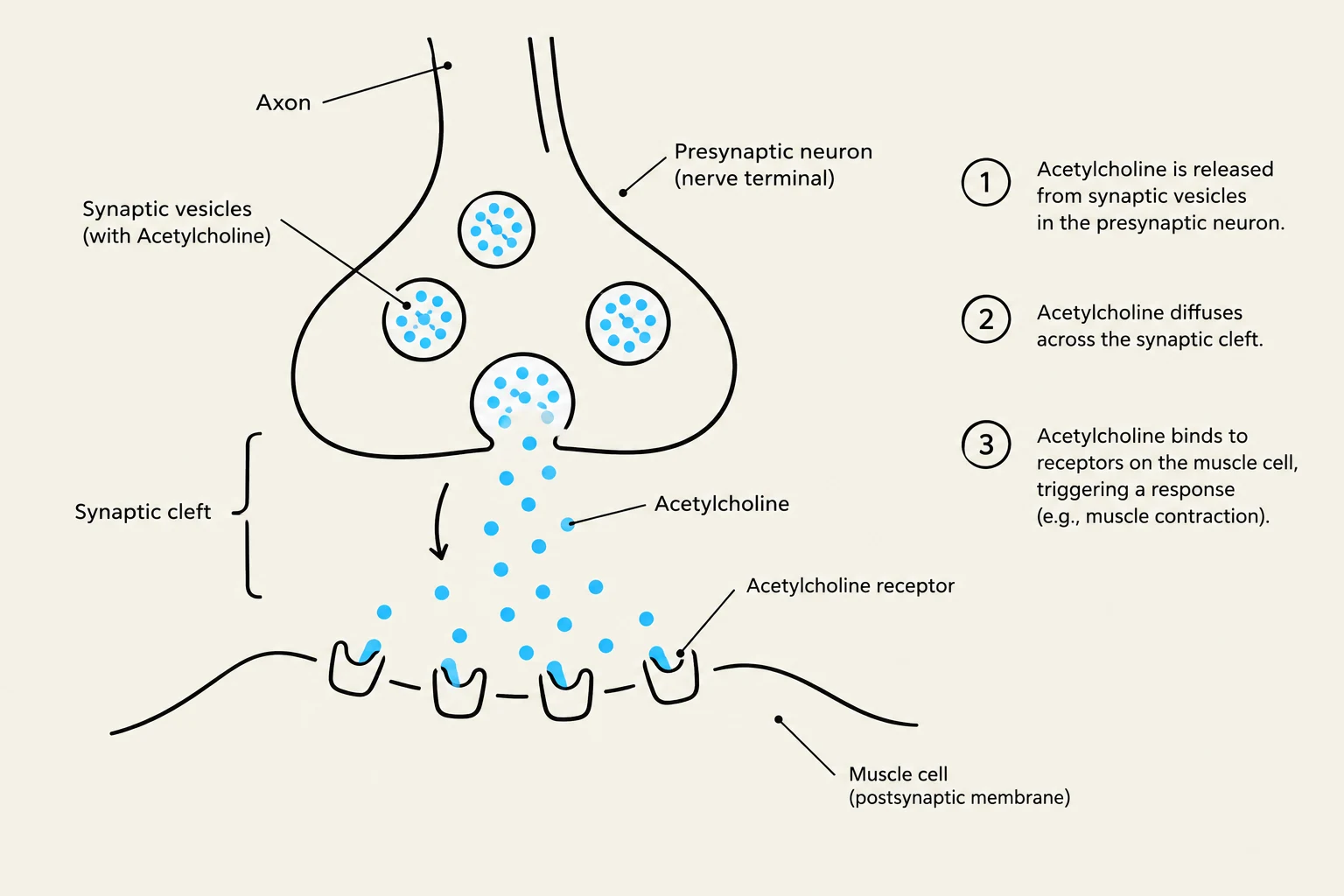
In a healthy system, the neurotransmitter acetylcholine (ACh) is released from the neuron and binds to nicotinic receptors on the muscle membrane to trigger contraction. In Myasthenia Gravis, the body's immune system produces acetylcholine receptor antibodies (specifically IgG antibodies).
These antibodies interfere with transmission in several ways:
They block the ACh receptors.
They decrease the total number of available receptors.
They physically destroy the receptor sites on the muscle membrane.
This process is often linked to abnormalities in the thymus gland; approximately 70–80% of patients have pathologic changes in the thymus, and 98% of those with a thymoma (a tumor of the thymus) test positive for these antibodies.
Clinical Presentation
The hallmark of MG is exertional weakness: muscle strength is relatively preserved at rest but worsens with activity and improves with rest.
Ocular/Bulbar Symptoms: Often the first signs (in 90% of cases), including ptosis (drooping eyelids) and diplopia (double vision). Patients may also experience difficulty chewing, swallowing (dysphagia), and speaking (dysarthria).
Generalized Weakness: Affects proximal muscles like the neck, shoulders, and hips.
Respiratory Risk: Involvement of the diaphragm can lead to hypoventilation and respiratory failure.
Diagram illustrating the process of synaptic transmission, where acetylcholine is released from a presynaptic neuron, diffuses across the synaptic cleft, and binds to receptors on a muscle cell, triggering a response like muscle contraction.
Medical Emergencies: Myasthenic vs. Cholinergic Crisis
Patients with MG must be monitored closely for two life-threatening crises:
Feature
Myasthenic Crisis
Cholinergic Crisis
Cause
Insufficient medication or drug resistance
Excess of cholinergic drugs (ACh excess)
Mechanism
Severe weakness leading to respiratory failure
Overstimulation of the Parasympathetic Nervous System
Key Signs
Quadriparesis, extreme difficulty swallowing
Miosis (constricted pupils), bradycardia, salivation, diarrhea, and muscle fasciculations
Clinical Note: Distinguishing between these two crises is vital. A myasthenic crisis requires more medication to improve strength, whereas a cholinergic crisis requires the immediate reduction of cholinergic drugs to prevent respiratory arrest from excessive secretions and bronchospasm.
Keep reading for free.
Create an account to finish this reading and make it part of your study routine.