Understanding Myocardial Infarction and Cardiovascular Disease
This content explains myocardial infarction (MI), a critical manifestation of cardiovascular disease. It details the pathophysiology from atherosclerosis to thrombus formation and obstruction, discusses modifiable and non-modifiable risk factors, outlines classic and atypical clinical presentations, and covers diagnost
Cardiovascular disease (CVD) stands as the leading cause of death for both men and women in the United States, contributing to approximately one in every four deaths. While many people live with chronic cardiovascular conditions for years due to advancements in medical technology and pharmacology, the most acute and life-threatening manifestation of this disease is the myocardial infarction (MI). Understanding the progression from arterial buildup to muscle death is essential for any healthcare professional.
Pathophysiology: From Plaque to Infarction
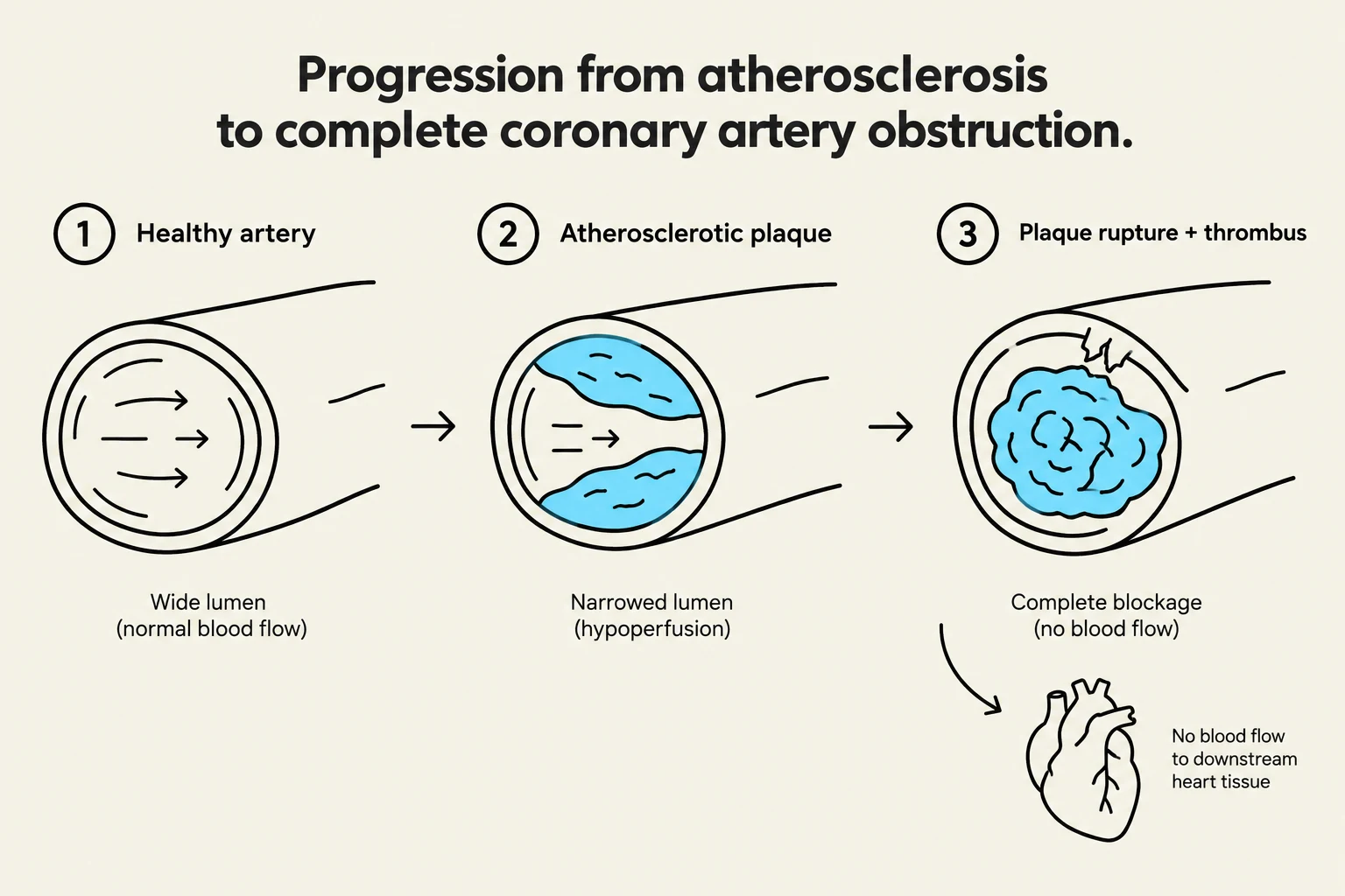
Diagram illustrating the progression of atherosclerosis leading to complete coronary artery obstruction, showing a healthy artery, an artery with an atherosclerotic plaque, and an artery with plaque rupture and thrombus, resulting in no blood flow to downstream heart tissue.
The journey toward a myocardial infarction typically begins with atherosclerosis. This is a chronic process involving the buildup of fatty, calcified plaques within the arterial walls, which leads to the hardening of the arteries.
As these plaques grow, they narrow the lumen of the coronary arteries, reducing the amount of oxygenated blood that can reach the heart muscle—a state known as hypoperfusion. The critical turning point occurs when one of these atherosclerotic plaques ruptures. This rupture triggers the body's clotting mechanism, leading to thrombus formation (a blood clot) at the site of the injury.
If the resulting thrombus is large enough, it causes a coronary artery obstruction. This blockage prevents blood from flowing to the downstream myocardium, resulting in:
Ischemia: A lack of oxygenated blood supply to the tissue.
Angina: Chest pain resulting from the heart muscle's inability to receive adequate perfusion.
Infarction: The actual destruction and death of the heart muscle tissue due to prolonged lack of oxygen.
Exam Tip: Remember the distinction between ischemia and infarction. Ischemia is the lack of blood flow (reversible if treated quickly), whereas infarction is the death of the tissue (permanent damage).
Risk Factors: Modifiable vs. Non-Modifiable
Identifying risk factors is the cornerstone of prevention. These are categorized into two distinct groups:
Modifiable Risk Factors
These are lifestyle choices or medical conditions that can be managed, treated, or reversed through intervention:
Cigarette Smoking: A major contributor to vascular damage.
Hypertension: High blood pressure that stresses arterial walls.
Dyslipidemia: Specifically high LDL (low-density lipoprotein) profiles.
Type 2 Diabetes: High blood glucose levels contribute to vascular complications.
Obesity and Sedentary Lifestyle: Lack of physical activity and excess weight increase cardiac strain.
Non-Modifiable Risk Factors
These are inherent characteristics that a patient cannot change:
Male Gender: Men are statistically at higher risk.
Postmenopausal Status: Female risk increases significantly after menopause.
Family History: A genetic predisposition to heart disease is a major red flag.
Clinical Manifestations and Symptom Variability
When an MI occurs, the clinical presentation can vary significantly. While some patients exhibit "textbook" symptoms, others may present in ways that are easily overlooked.
Classic Symptoms
The hallmark signs of an acute MI include:
Substernal Chest Pain: Often described as heavy or crushing.
Referred Pain: Pain that radiates to the shoulder, down the arm, to the jaw, teeth, or the back.
Dyspnea: Shortness of breath.
Diaphoresis: Profuse, cold sweating.
Gastrointestinal Distress: Nausea and vomiting.
Psychological Signs: Patients frequently report a "feeling of impending doom," a sense that something is profoundly wrong.
Atypical Presentations: The "Silent" Risk
Clinicians must exercise high suspicion when treating specific populations, as they may not present with classic chest pain:
Geriatric Patients: Older adults may present with atypical symptoms or may be entirely asymptomatic.
Female Patients: Women are more likely to experience indigestion, extreme fatigue, or pain in the jaw and neck rather than standard chest pressure.
Diabetic Patients: These individuals may experience "silent symptoms," where the lack of typical pain makes the MI difficult to detect.
Clinical Pearl: In these high-risk groups, do not wait for chest pain to occur. Rely on physical assessments, such as monitoring blood pressure, checking oxygen saturation (SpO2), and auscultating heart sounds, to detect signs of cardiac distress.
Diagnostics and Immediate Interventions
Comparison of normal EKG and ST-elevation myocardial infarction (STEMI).
In the emergency setting, time is muscle. The goal is to restore perfusion as quickly as possible to limit the extent of myocardial destruction.
Diagnostic Gold Standards
EKG (Electrocardiogram): This is the gold standard for diagnosing an MI. It allows clinicians to differentiate between a STEMI (ST-Elevation Myocardial Infarction), which indicates significant injury, and an NSTEMI (Non-ST Elevation Myocardial Infarction), which typically shows ST depression or T-wave inversion indicating ischemia.
Cardiac Biomarkers: Blood tests are used to detect proteins released when heart muscle is damaged. Key markers include:
Troponin: Highly sensitive and specific to cardiac injury.
CK-MB (Creatine Kinase-Myocardial Band): Very specific to cardiac tissue.
BNP (B-type Natriuretic Peptide): Often used to correlate with heart failure.
Emergency Interventions
The "Door-to-Balloon" time—the time from when a patient enters the hospital to when a coronary artery is opened via angioplasty—should ideally be within 90 minutes.
Coronary Angioplasty (PCI): A catheter is threaded through the femoral or radial artery to the site of the obstruction. A balloon is inflated to compress the plaque against the vessel walls, and a stent is often placed to keep the artery open.
Pharmacological Support:
Aspirin: Given as a chewed dose (e.g., 325 mg) to ensure rapid absorption; it acts as an antiplatelet to prevent the clot from growing larger.
Nitroglycerin: Dilates coronary arteries to increase blood flow; typically administered up to 3 doses, 5 minutes apart.
Morphine/Fentanyl: Used for pain management, though clinicians must be cautious as morphine can potentially mask symptoms or worsen the MI in some contexts.
Post-Intervention and Long-Term Care
Diagram illustrating the process of coronary angioplasty with balloon inflation and stent placement to open a narrowed artery.
Following an acute event or surgical intervention like a CABG (Coronary Artery Bypass Grafting), the focus shifts to recovery and prevention. Patients must be educated on medication adherence, the importance of cardiac rehabilitation, and lifestyle modifications such as the DASH diet and regular exercise.
For those recovering from invasive procedures, nurses must remain vigilant for complications such as infection (indicated by purulent drainage or fever) or bleeding, especially if the patient is on anticoagulation therapy.
Keep reading for free.
Create an account to finish this reading and make it part of your study routine.