Understanding Musculoskeletal Injuries and Fracture Classifications
This content provides a comprehensive overview of musculoskeletal injuries, including fractures, dislocations, sprains, and strains. It details various fracture classifications based on completeness, exposure, and pattern, and explains the stages of bone healing and potential complications.
The musculoskeletal system provides the structural framework for the human body, enabling movement, protection of vital organs, and support. However, this system is susceptible to various injuries ranging from minor soft tissue strains to severe bone fractures and dislocations. Understanding the nuances of these injuries—how they are classified, how the body heals them, and the potential complications that arise—is essential for clinical assessment and effective patient care.
Overview of Musculoskeletal Injuries
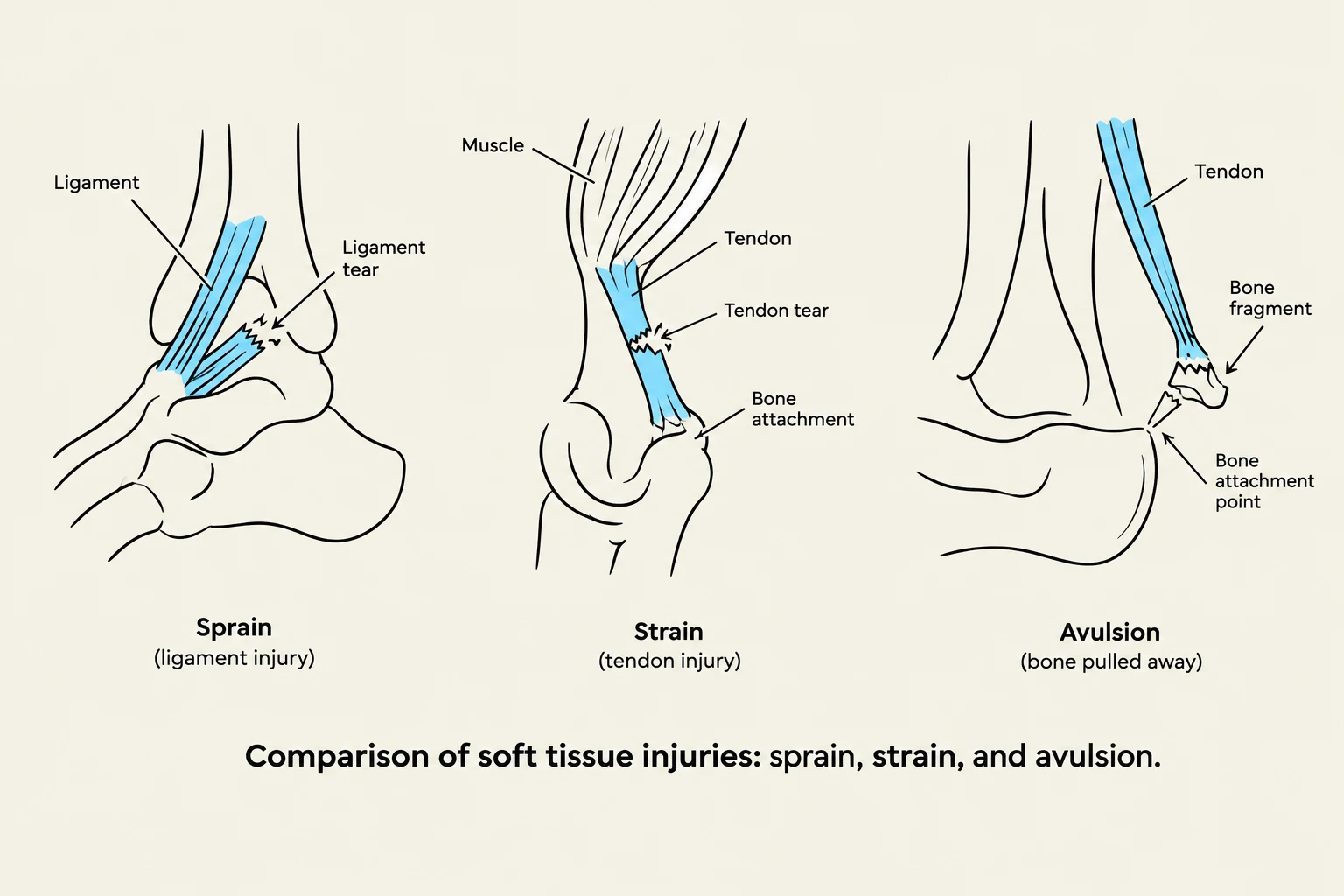
Comparison of soft tissue injuries: sprain, strain, and avulsion.
Musculoskeletal injuries are broadly categorized into those affecting the bones and those affecting the supporting soft tissues.
Fractures: A break in the continuity of a bone that occurs when the force applied to the bone exceeds its compressive strength.
Dislocations: The temporary displacement of a bone from its normal position within a joint. A partial dislocation is specifically referred to as a subluxation.
Sprains: An injury involving a tear or damage to a ligament.
Strains: An injury involving a tear or damage to a tendon.
Avulsion: A specific type of injury where a tendon or ligament is completely separated from its attachment point on the bone.
Fracture Classifications
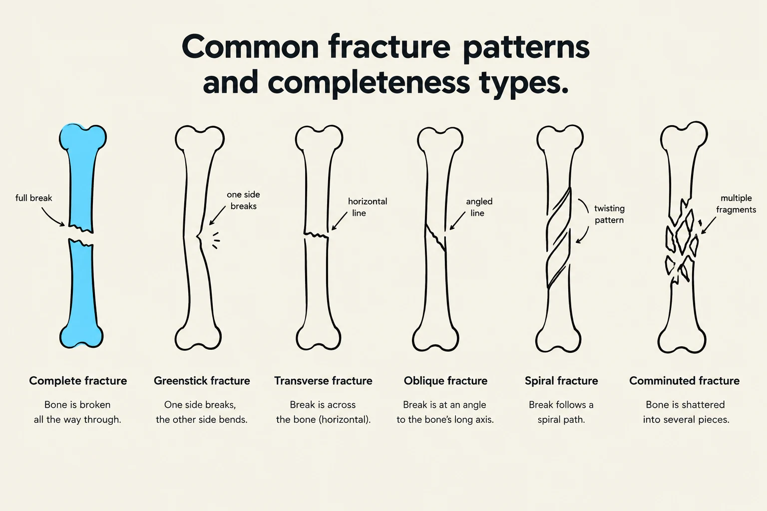
This diagram illustrates common fracture patterns and completeness types in bones, including complete, greenstick, transverse, oblique, spiral, and comminuted fractures.
Because no two breaks are identical, clinicians use several classification systems to describe the nature of a fracture. This helps in determining the appropriate treatment, such as whether a patient requires a cast, traction, or surgical intervention like Open Reduction Internal Fixation (ORIF).
Completeness and Exposure
Fractures are first categorized by how much of the bone is affected and whether the injury reaches the surface of the skin:
Complete vs. Incomplete: A complete fracture involves a full break through the bone. An incomplete fracture does not extend through the entire bone. Common incomplete types found in pediatric patients include:
Greenstick fractures: Where the bone bends and cracks but does not break through.
Torus fractures: Often described as a "bulging" fracture.
Bowing fractures: Common in children, these often involve a pair of bones (like the radius-ulna or tibia-fibula). One bone may fracture completely, which prevents the second bone from breaking, causing it to bow instead.
Open vs. Closed: An open (compound) fracture occurs when the break communicates with the external environment, creating a risk of infection. A closed (simple) fracture does not break the skin.
Fracture Patterns
The geometry of the break is described by its pattern:
Linear: A break along the long axis of the bone.
Oblique: An angled break.
Spiral: A break that twists around the bone.
Transverse: A horizontal break perpendicular to the bone's axis.
Comminuted: A severe break where the bone is shattered into multiple fragments.
Special Fracture Types
Certain fractures are categorized by the underlying cause or the specific tissue involved:
Pathologic Fractures: These occur at sites of pre-existing abnormalities, such as tumors or osteoporosis.
Stress Fractures: These result from repeated stress applied to normal or abnormal bone.
Transchondral Fractures: These involve the separation of the articular cartilage from the main shaft of the bone and are frequently seen in the ankle, elbow, knee, or wrist.
Exam Tip: When assessing a patient, remember the "5 P's" of neurovascular assessment to check for complications: Pain, Pulses, Pallor, Paralysis, and Paresthesia.
The Physiology of Fracture Healing
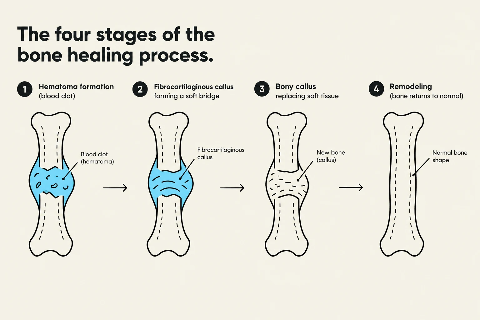
The four stages of bone healing: hematoma formation, fibrocartilaginous callus, bony callus, and remodeling.
The body undergoes a complex, four-stage process to repair broken bone:
Hematoma Formation: Immediately following the injury, bleeding occurs at the fracture site. This leads to vasoconstriction and the formation of a hematoma (blood clot). As the clot stabilizes, granulation tissue begins to form, and osteoblasts and osteoclasts are recruited to the site.
Fibrocartilaginous Callus Formation: Fibroblasts and osteoblasts begin the reconstruction process, creating a bridge of fibrocartilage between the broken ends.
Bony Callus Formation: The fibrocartilaginous callus is gradually replaced by mature bone. During this stage, any excess callus is reabsorbed by the body.
Remodeling Phase: This is the final, long-term stage where necrotic (dead) bone is removed, and compact bone replaces spongy bone to restore the bone's original strength and shape.
Clinical Manifestations and Risk Factors
Recognizing a Fracture
Clinicians look for several key signs when a fracture is suspected:
Pain and swelling at the site.
Visible deformity or misalignment.
Numbness (indicating nerve involvement).
Evidence of internal or external blood loss.
Confirmation via diagnostic imaging such as X-ray, CT, or MRI.
Risk Factors
The likelihood of sustaining a fracture varies by demographic:
Age: Younger clients are more prone to sports-related injuries, while older adults are at higher risk due to falls and bone diseases.
Bone Disease: Conditions like osteoporosis weaken the bone structure.
Nutrition: Poor nutritional status can compromise bone density and healing.
Complications of Fracture Healing
If the healing process is interrupted or goes wrong, several complications can arise:
Nonunion: The bone ends fail to grow together, and the gap is filled with fibrous tissue instead of bone.
Delayed Union: The healing process takes significantly longer than the expected 8–9 months.
Malunion: The bone heals, but in an improper or deformed position.
Beyond healing issues, acute complications can be life-threatening:
Compartment Syndrome: Swelling or bleeding within a muscle compartment increases pressure on capillaries and nerves, potentially causing muscle damage.
Deep Vein Thrombosis (DVT): A blood clot in a deep vein, often requiring anticoagulants.
Fat Embolism Syndrome: Fat from the bone marrow enters the bloodstream, which can lead to respiratory failure or neurological dysfunction.
Infection: Particularly common in open fractures, often caused by organisms like Staphylococcus or Pseudomonas.
Keep reading for free.
Create an account to finish this reading and make it part of your study routine.