Understanding Muscle Mechanics, Types, and Anatomy
An in-depth explanation of the muscular system, covering the mechanics of muscle contraction, the three types of muscle tissue (smooth, skeletal, and cardiac), muscle roles in joint movement, and how skeletal muscles are named and anatomically structured. It also discusses muscle growth through hypertrophy and atrophy.
The muscular system is a complex network of tissues responsible for nearly every movement your body performs, from the rapid beating of your heart to the controlled lifting of a heavy weight. While you are born with a fixed number of muscle cells, the size and strength of these cells can change significantly based on how you use them. Understanding how muscles function requires looking at their mechanical principles, the different types of tissue that make up the system, and the specific ways they are named and organized in the human body.
The Mechanics of Contraction
A fundamental rule of kinesiology is that muscles can only pull; they cannot push. Because muscles work by shortening, they must operate in pairs to facilitate movement in multiple directions. For example, to flex your arm, your biceps contract, but to extend it, a different muscle (the triceps) must take over.
The All or None Principle
When a muscle is stimulated, it does so through "motor units," which consist of a nerve and the muscle fibers it controls. The All or None Principle states that a single motor unit will either contract to its fullest potential or not at all; there is no such thing as a "partial" contraction for an individual motor unit. The overall strength (or grade) of a muscle contraction depends on how many of these motor units are activated at once.
Three Types of Contractions
Depending on the relationship between the muscle's strength and the load it is moving, contractions are categorized into three types:
Concentric Contraction: The muscle shortens, and the insertion (the movable end) moves closer to the origin (the stable end). An example is lifting a heavy book toward your shoulder.
Eccentric Contraction: The muscle lengthens while it is still contracting. This often happens when the load is greater than the muscle's strength, such as when you are slowly lowering a heavy object.
Isometric Contraction: The muscle tension increases, but there is no change in the length of the muscle or the angle of the joint. Common examples include holding a plank or performing a wall sit.
Study Tip: Origin vs. Insertion
To remember the difference, think of the Origin as the "anchor"—it is the most proximal, least moveable part of the muscle. The Insertion is the "active" end—it is the most distal, most moveable part that pulls on the bone to create movement.
Muscle Roles in Joint Movement
No single muscle acts entirely alone during movement. Instead, muscles work in specialized roles to ensure stability and efficiency:
Agonist (Prime Mover): The muscle primarily responsible for producing a specific movement.
Antagonist: The muscle that opposes the movement of the agonist. To allow the agonist to move, the antagonist must generally relax.
Fixator: A muscle that contracts to hold the bone of origin steady, providing a stable base so the bone of insertion can move effectively.
The Three Types of Muscle Tissue
The human body utilizes three distinct types of muscle tissue, each specialized for different physiological needs:
Muscle Type
Striated?
Control
Location
Primary Function
Smooth
No
Involuntary
Hollow organs, blood vessels
Internal movement of fluids (blood, waste)
Skeletal
Yes
Voluntary
Attached to bones
Locomotion, breathing, posture
Cardiac
Yes
Involuntary
Heart only
Pumping blood via internal contraction system
Smooth muscle is non-striated and manages the movement of products through the body, such as food through the digestive tract. Skeletal muscle is striated and allows for voluntary external movements. Finally, cardiac muscle is unique; while it is striated like skeletal muscle, it contains gap junctions between cells. These gaps allow electrical signals to pass quickly between cells, ensuring the heart contracts in a coordinated, rhythmic squeeze.
Functions of the Muscular System
The muscular system serves three vital roles for survival and daily activity:
Locomotion: This includes external movements like running or throwing, as well as internal movements like the beating of the heart or the movement of food through the intestines.
Posture: Muscles like the erector spinae work constantly to maintain body position, such as keeping your neck and spine upright.
Heat Production: Muscle contraction produces heat as a byproduct. This is essential for maintaining a basal body temperature of approximately 36.5∘C. When you are cold, your body triggers shivering—involuntary muscle contractions—to generate heat and raise your temperature.
Naming and Anatomy of Skeletal Muscles
Skeletal muscles are named using several standardized criteria, which helps anatomists identify them quickly:
Location: Named after the bone they are near (e.g., Temporalis overlies the temporal bone).
Shape: Named for distinctive forms (e.g., Deltoid is triangular; Trapezius is trapezoid-shaped).
Size: Named by relative size (e.g., Gluteus maximus vs. minimus).
Direction of Fibers: (e.g., Rectus for straight, Transverse for right angles).
Number of Origins: (e.g., Biceps meaning two heads).
Action: Named for the movement they produce (e.g., Adductor moves a limb toward the midline).
Key Muscle Examples
Below are several major muscles, their origins, insertions, and primary actions:
The Upper Body
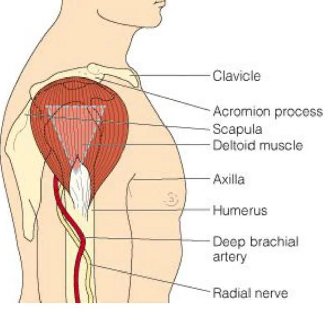
Deltoid: Originates on the clavicle and scapula; inserts on the humerus. It is responsible for shoulder flexion, extension, rotation, and abduction.
An illustration of the human shoulder and upper arm, labeling the clavicle, acromion process, scapula, deltoid muscle, axilla, humerus, deep brachial artery, and radial nerve.
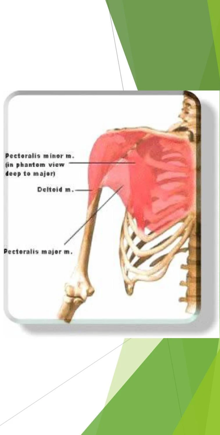
Pectoralis Major: Originates on the clavicle, sternum, and ribs 2-6; inserts on the humerus. It assists in shoulder flexion and abduction.
An illustration of the human pectoral muscles and deltoid muscle.
Biceps: Originates on the scapula and inserts on the proximal radius. It facilitates elbow flexion and supination of the hand.
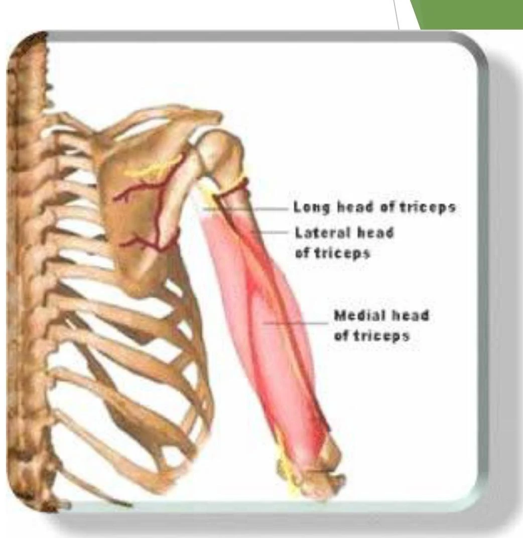
Triceps Brachii: Originates on the scapula and humerus; inserts on the proximal ulna. It is the primary muscle for elbow extension.
An illustration of the triceps brachii muscle, showing its three heads: long, lateral, and medial.
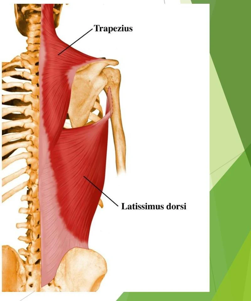
Trapezius: Originates on the occipital bone; inserts on the clavicle and scapula. It assists in shoulder elevation and retraction.
An illustration of the trapezius and latissimus dorsi muscles.
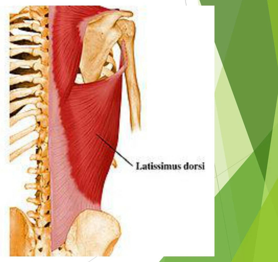
Latissimus Dorsi: Originates on the vertebrae, sacrum, ilium, and ribs; inserts on the humerus. It performs shoulder extension, adduction, and medial rotation.
An illustration of the latissimus dorsi muscle.
The Core and Lower Body
Abdominals: Originate on the pubic crest/symphysis and insert on the xiphoid process and inferior ribs. They facilitate flexion of the waist.
Quadriceps: A group of muscles (including Rectus Femoris and Vastus muscles) that insert into the patella and tibia to perform knee extension.
An illustration of the quadriceps femoris muscles of the human thigh, showing the vastus lateralis, vastus intermedius, vastus medialis, and rectus femoris.
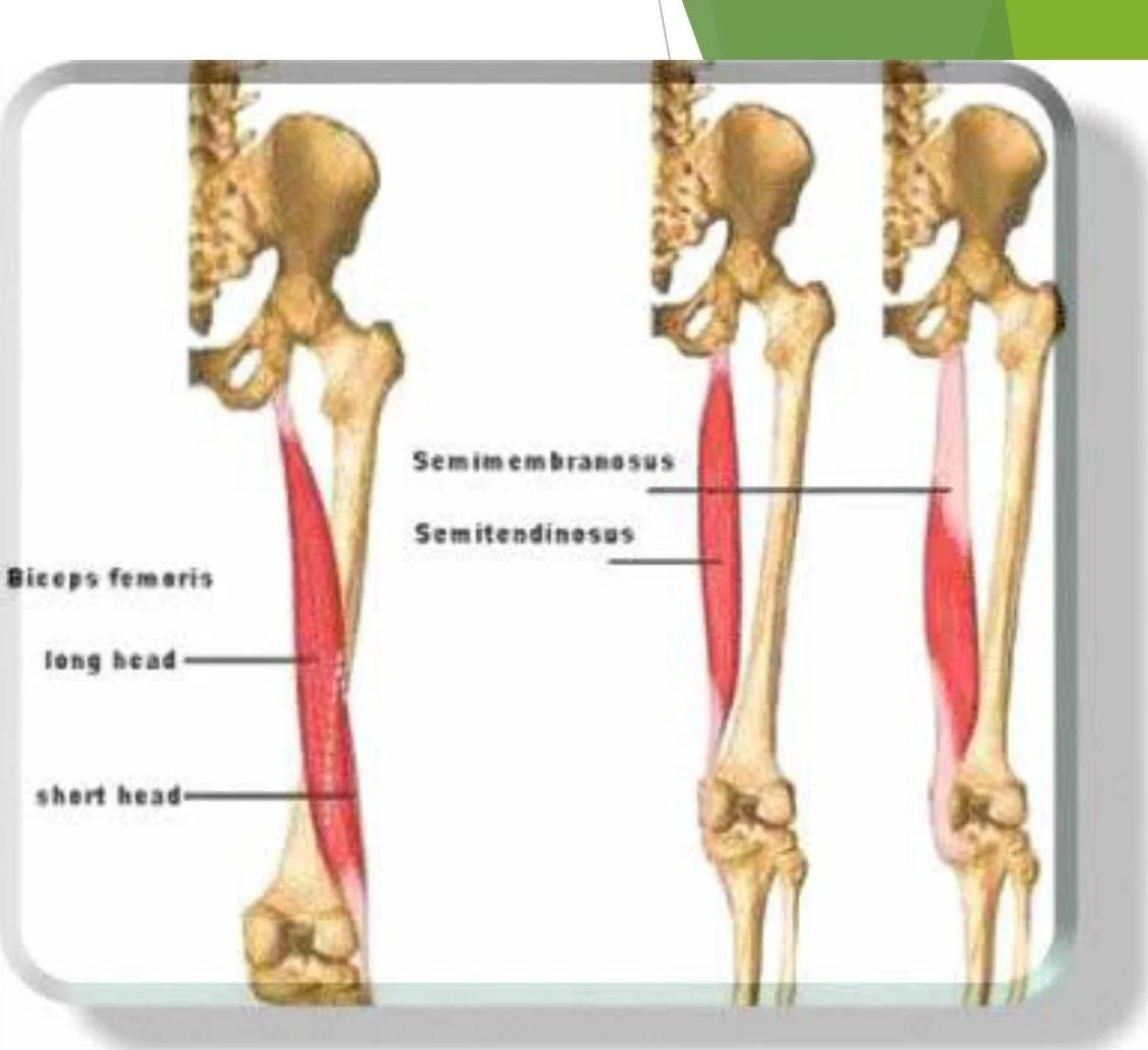
Hamstrings: A group of muscles (Biceps Femoris, Semitendinosus, Semimembranosis) that perform knee flexion.
An illustration of the hamstring muscles, including the biceps femoris, semimembranosus, and semitendinosus, attached to the femur and tibia.
Gluteus Maximus: Originates on the iliac crest, sacrum, and coccyx; inserts on the IT band and femur. It is responsible for hip extension and abduction.
Muscle Growth: Hypertrophy vs. Atrophy
While you cannot increase the number of muscle cells you have, you can change their size through use.
Hypertrophy: This is the increase in the size of existing muscle cells. When you subject a muscle to increased loads (such as weightlifting), your body responds by adding more muscle tissue (actin and myosin) to meet the demand.
Atrophy: This is the decrease in muscle cell size due to inactivity or injury. This can occur in sedentary individuals, people in casts, or even astronauts in space due to the lack of gravity.
To maintain muscle mass and prevent atrophy, staying active is essential.
Keep reading for free.
Create an account to finish this reading and make it part of your study routine.