


The middle ear and the surrounding temporal bone are complex structures essential for hearing and balance. When infections, structural changes, or abnormal growths occur in these areas, the clinical implications can range from mild hearing loss to life-threatening neurological complications. This article explores three distinct but clinically significant conditions: mastoiditis, cholesteatoma, and otosclerosis.
Mastoiditis: Infection and Complications
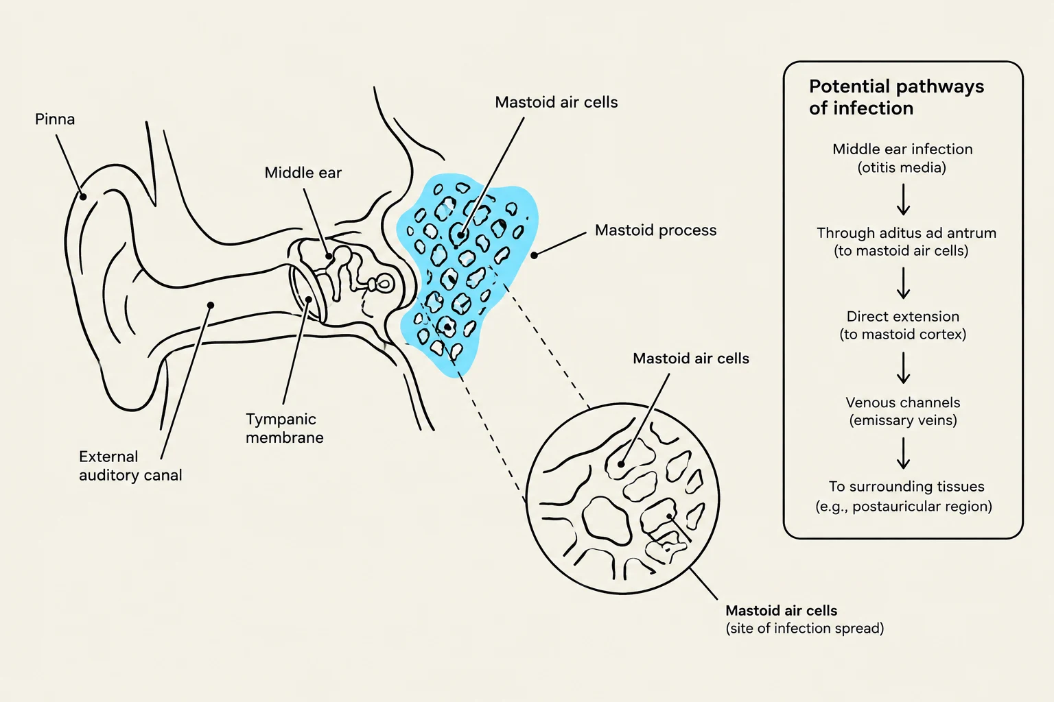
Mastoiditis is a serious inflammatory condition involving the mastoid process, which is the part of the temporal bone containing numerous air cells that communicate with the middle ear space. It most commonly occurs as a complication of acute otitis media (AOM) when the infection spreads from the middle ear into these air cells.
Clinical Presentation and Diagnosis
Patients with mastoiditis often present with a spiking fever occurring weeks after an inadequately treated episode of AOM. Physical examination typically reveals:
- Mastoid tenderness (often described as a "doughy" feeling).
- Retroauricular edema (swelling behind the ear), erythema, and heat.
- A protruding auricle and the loss of the normal postauricular crease.
The diagnostic study of choice is a CT scan, which will show the coalescence of the mastoid air cells due to the destruction of their bony septa.
Exam Tip: Differential Diagnosis In clinical practice, it is vital to distinguish true mastoiditis from "pseudo-mastoiditis." Other conditions that can mimic the presentation of mastoid swelling include secondary cellulitis from otitis externa, parotitis, or cervical lymphadenitis.
Intracranial Extension
The most critical concern with mastoiditis is its potential for intracranial extension. Because the infection is located within the temporal bone, it can breach the skull base, leading to:
Keep reading for free.
Create an account to finish this reading and make it part of your study routine.
Continue reading freeAlready have an account? Log in
Updated