


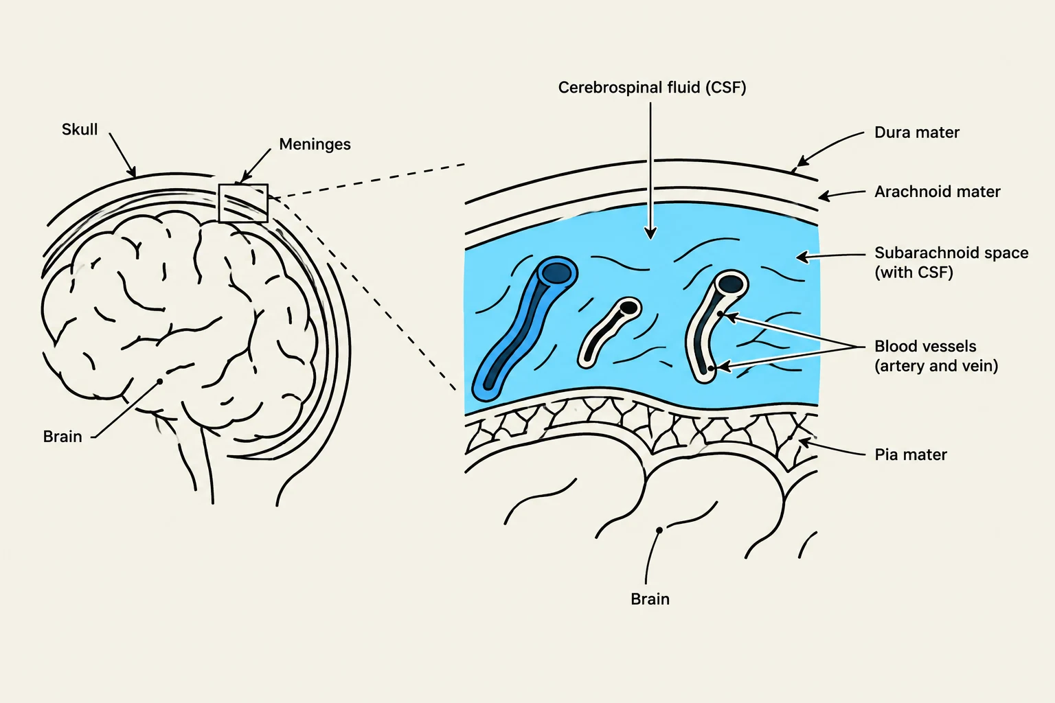
The central nervous system (CNS) is not merely floating loosely within the skull; it is protected by a sophisticated, multi-layered system of membranes known as the meninges. These layers serve as physical barriers, provide structural support, and facilitate the circulation of cerebrospinal fluid (CSF). For students of anatomy and clinical medicine, the ability to differentiate between the various "spaces" created by these layers is critical, as many life-threatening pathologies—such as hemorrhages—are defined specifically by which space they occupy.
The Anatomy of the Meninges
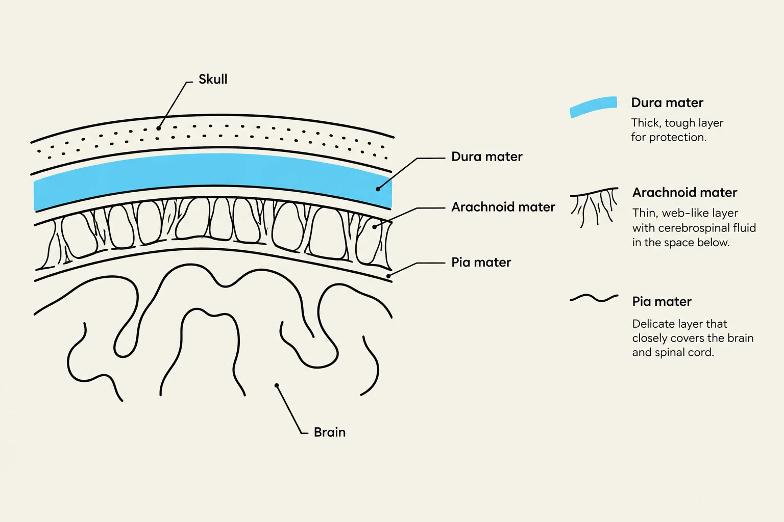
To understand the spaces, one must first understand the layers that define them. The meninges consist of three distinct membranes that wrap around the brain and spinal cord.
- Dura Mater: The outermost, toughest, and most fibrous layer. It provides a heavy-duty protective shell.
- Arachnoid Mater: The middle layer, characterized by a web-like appearance. It is much thinner and more delicate than the dura.
- Pia Mater: The innermost layer, which is highly vascularized and adheres directly to the surface of the brain and spinal cord, following every contour and sulcus.
The Epidural Space: The Potential Space
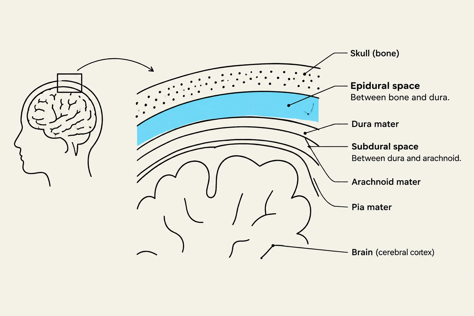
The epidural space is located between the inner surface of the skull and the outer surface of the dura mater.
In the cranium, the epidural space is often considered a "potential space." Under normal physiological conditions, there is no actual gap; the dura is pressed firmly against the bone. However, if a blood vessel (most commonly the middle meningeal artery) ruptures, blood accumulates in this area, turning the potential space into a real, expanding space.
Keep reading for free.
Create an account to finish this reading and make it part of your study routine.
Continue reading freeAlready have an account? Log in
Updated