A comprehensive overview of liver anatomy, physiology, types of liver failure, classification of cirrhosis, clinical manifestations, complications, diagnostic assessments, and management strategies for liver disease.
The liver is a powerhouse of metabolic activity, acting as a central hub for detoxification, nutrient processing, and systemic regulation. When this vital organ is subjected to chronic injury or sudden insult, its ability to maintain homeostasis collapses, leading to life-threatening conditions. Understanding the transition from healthy liver function to the irreversible scarring of cirrhosis is essential for clinical practice.
Liver Anatomy and Physiology
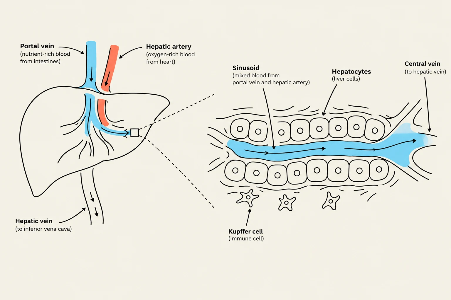
The liver is a complex organ composed of functional units called lobules. Blood enters the liver through two primary sources: the hepatic artery, which provides oxygenated blood, and the hepatic portal vein, which brings nutrient-rich, deoxygenated blood from the gastrointestinal tract. These vessels merge into sinusoids—specialized capillaries lined with hepatocytes—where exchange occurs.
To remember the diverse roles of the liver, use the mnemonic P.D.S.M.:
P - Production: The liver synthesizes Albumin (maintaining osmotic pressure), Bile (for fat digestion), and essential Coagulation factors (to prevent bleeding).
D - Detoxification: It filters chemicals, drugs, and toxins from the blood. A critical process is converting highly toxic ammonia into urea, which can then be excreted by the kidneys.
S - Storage: It acts as a reservoir for Glycogen (for glucose regulation) and vitamins A, D, E, K, and B12.
M - Metabolism: It breaks down proteins and amino acids to fuel the body.
Diagram illustrating blood flow through the liver, showing the portal vein, hepatic artery, sinusoids, hepatocytes, Kupffer cells, and central vein.
Types of Liver Failure
Liver failure is clinically categorized by its speed of onset and the underlying pathology.
Acute Liver Failure (Fulminant)
Acute failure is characterized by a rapid onset of symptoms, often occurring within weeks of an insult. The most common cause in the United States is Acetaminophen overdose. Other causes include viral hepatitis and certain metabolic disorders.
Chronic Liver Failure and Cirrhosis
Chronic failure is a progressive process. Over time, repeated injury leads to Cirrhosis, which is defined by extensive, irreversible scarring (fibrosis) and the destruction of healthy hepatocytes. In cirrhosis, the liver's architecture is replaced by regenerative nodules—clumps of living cells surrounded by bands of non-functional scar tissue.
Exam Tip: If a question asks about the difference between acute and chronic failure, remember that "Fulminant" refers to the rapid, acute onset, while "Cirrhosis" is the end-stage result of chronic, progressive damage.
Classifying Cirrhosis
The type of cirrhosis is determined by its primary cause:
Postnecrotic Cirrhosis: Results from viral hepatitis, drugs, or toxins.
Laennec’s Cirrhosis: Specifically caused by chronic alcohol consumption (ETOH).
Biliary Cirrhosis: Results from chronic biliary obstruction or autoimmune diseases.
The severity of the disease is measured in two stages:
Compensated Stage: The liver is scarred, but it can still perform enough essential functions to maintain relatively stable physiology.
Decompensated Stage: The liver can no longer meet the body's needs. The organ may shrink in size, and severe clinical manifestations emerge.
Clinical Manifestations and Physical Assessment
As the liver fails, the body exhibits a wide range of systemic signs. These can be categorized by the body system they affect:
Integumentary and Visual
Jaundice/Icterus: Yellowing of the skin and eyes due to the buildup of unconjugated bilirubin.
Spider Angiomas & Palmar Erythema: Red, spider-like vessels on the skin and reddened palms, often caused by increased estrogen levels that the liver can no longer metabolize.
Ecchymosis & Petechiae: Easy bruising or small red spots due to a lack of clotting factors.
Gastrointestinal and Cardiovascular
Ascites: The accumulation of protein-rich fluid in the peritoneal cavity.
Fetor Hepaticus: A distinct, musty breath odor caused by the buildup of toxins.
Edema: Peripheral swelling in the extremities.
Neurological
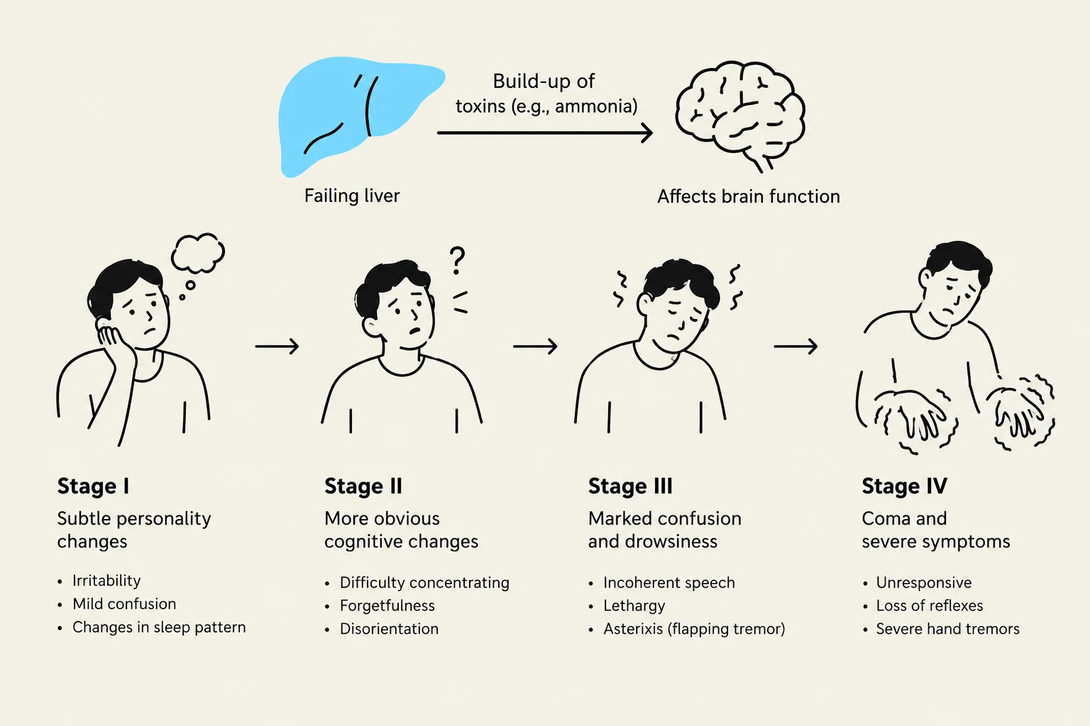
Hepatic Encephalopathy (HE): A complex cognitive syndrome caused by toxins (like ammonia) reaching the brain.
Stage I: Personality changes, emotional lability, and sleep disturbances.
Stage II: Mental confusion and Asterixis (a "flapping" hand tremor when arms are outstretched).
Stage III: Marked confusion, stupor, and hyperreflexia.
Stage IV: Unresponsiveness, coma, and seizures.
This diagram illustrates the four stages of hepatic encephalopathy, a neurological complication of liver disease, showing the progression of symptoms from subtle personality changes to coma and severe symptoms due to the build-up of toxins affecting brain function.
Complications of Cirrhosis
The most significant complication of cirrhosis is Portal Hypertension—an increase in pressure within the portal vein system. Because blood follows the path of least resistance, it shunts away from the liver into collateral vessels, causing them to become dilated and engorged.
This leads to several dangerous conditions:
Esophageal Varices: Dilated veins in the esophagus that are prone to life-threatening hemorrhage.
Splenomegaly: An enlarged spleen caused by blood backing up from the liver.
Caput Medusae: Visible, dilated veins radiating from the umbilicus.
Hemorrhoids: Dilated veins in the rectum.
Spontaneous Bacterial Peritonitis (SBP): A life-threatening infection of the ascitic fluid.
Hepatorenal Syndrome (HRS): Kidney failure that follows liver failure, often indicating a very poor prognosis.
Laboratory and Diagnostic Assessment
To evaluate liver health, clinicians rely on a combination of blood work and imaging.
Laboratory Indicators
Liver Enzymes: Elevated AST, ALT, and LDH indicate hepatocellular injury.
Bilirubin: Elevated levels lead to jaundice.
Ammonia: Elevated levels are a primary driver of encephalopathy.
Coagulation & Proteins: Decreased Albumin and clotting factors lead to a prolonged PT/INR (increased bleeding risk).
Hematology: Thrombocytopenia (low platelets) is common.
Diagnostic Procedures
Liver Biopsy: The "gold standard" for diagnosing cirrhosis, as it allows for direct microscopic examination of tissue.
Imaging: Ultrasound, MRI, and X-rays to visualize liver size and structure.
Endoscopy (EGD/ERCP): Used to visualize esophageal varices or biliary obstructions.
Management and Treatment
Because the scarring in cirrhosis is irreversible, treatment focuses on managing symptoms and preventing further damage.
Address the Underlying Cause: This includes stopping alcohol consumption or starting antiviral therapy for Hepatitis B or C.
Manage Ascites: Treatment includes low-sodium diets, diuretics (like Spironolactone), and paracentesis (draining fluid from the abdomen).
Prevent Variceal Bleeding: Beta-blockers (like Propranolol) are used to decrease blood pressure in the portal system.
Treat Encephalopathy:Lactulose is used to promote the excretion of ammonia through the stool.
Liver Transplantation: For patients with end-stage liver failure, a transplant is often the only definitive treatment.
Clinical Note: When managing a patient with ascites, always measure the abdominal girth at the umbilicus at the end of exhalation to ensure consistent monitoring.
Keep reading for free.
Create an account to finish this reading and make it part of your study routine.